. 2009 Mar 19:10:115.
doi: 10.1186/1471-2164-10-115.

Independent evolution of neurotoxin and flagellar genetic loci in proteolytic Clostridium botulinum

Affiliations

Independent evolution of neurotoxin and flagellar genetic loci in proteolytic Clostridium botulinum

Andrew T Carter et al. BMC Genomics. .

Abstract

Background: Proteolytic Clostridium botulinum is the causative agent of botulism, a severe neuroparalytic illness. Given the severity of botulism, surprisingly little is known of the population structure, biology, phylogeny or evolution of C. botulinum. The recent determination of the genome sequence of C. botulinum has allowed comparative genomic indexing using a DNA microarray.

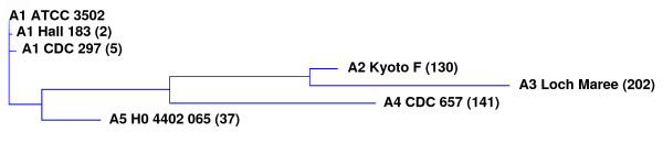
Results: Whole genome microarray analysis revealed that 63% of the coding sequences (CDSs) present in reference strain ATCC 3502 were common to all 61 widely-representative strains of proteolytic C. botulinum and the closely related C. sporogenes tested. This indicates a relatively stable genome. There was, however, evidence for recombination and genetic exchange, in particular within the neurotoxin gene and cluster (including transfer of neurotoxin genes to C. sporogenes), and the flagellar glycosylation island (FGI). These two loci appear to have evolved independently from each other, and from the remainder of the genetic complement. A number of strains were atypical; for example, while 10 out of 14 strains that formed type A1 toxin gave almost identical profiles in whole genome, neurotoxin cluster and FGI analyses, the other four strains showed divergent properties. Furthermore, a new neurotoxin sub-type (A5) has been discovered in strains from heroin-associated wound botulism cases. For the first time, differences in glycosylation profiles of the flagella could be linked to differences in the gene content of the FGI.

Conclusion: Proteolytic C. botulinum has a stable genome backbone containing specific regions of genetic heterogeneity. These include the neurotoxin gene cluster and the FGI, each having evolved independently of each other and the remainder of the genetic complement. Analysis of these genetic components provides a high degree of discrimination of strains of proteolytic C. botulinum, and is suitable for clinical and forensic investigations of botulism outbreaks.


Figures

Figure 1
Figure 1
Whole genome analysis of 61 strains of proteolytic C. botulinum and C. sporogenes. Each row of the heatmap represents a strain (indicated at right), and its branch on the dendrogram is coloured according to type of neurotoxin formed (indicated at left of heatmap; spo refers to C. sporogenes). Although lost at this resolution, each microarray probe is represented by a vertical column within this row, from left to right first the 19 probes for each CDS of ATCC 3502 plasmid pBOT3502, followed by probes for chromosomal CDSs, from CBO3648 to CBO001. The colour of each column in the heatmap is an indicator of test signal over reference (ATCC 3502) signal channel ratio. Yellow columns represent probes which hybridised to both test and reference isolates equally, those in blue hybridised more strongly to the reference strain, and those in red hybridised more strongly to the test strain. Microarray features with fluorescent signals lower than 100 units (background noise), plus those CDSs not represented on the microarray are coloured grey. Distance measurements between 0 and 1.0 are indicated in the non-linear scale underneath the dendrogram. Clades 1 to 9 (brackets at right), are groups of strains which cluster at a distance measurement value of 0.3. The four main regions of variability (clusters of blue-coloured columns) are CDSs associated with pBOT3502, the Flagellar Glycosylation Island (FGI), and the two prophages, Φ-CB1 and Φ-CB2 (indicated above heatmap).
Figure 2
Figure 2
Heatmap comparison of CDSs in the Flagellar Glycosylation Island (FGI). Top: See Figure 1 for explanation of heatmap format. Strains are ordered by FGI CDS data; dendrogram and strain names at right are coloured (as for Figure 1) for type of neurotoxin formed. Filled triangles (bottom of heatmap): approximately 10 CDS intervals (data for some CDSs are absent). CDSs of FGI-I (CBO2666 – CBO2692) are highly conserved and those of FGI-II (CBO2696 – CBO2729) are less so. Hybridisation profiles divided strains into 6 divisions, numbered at left. Mass of glycans detected by mass spectrometry analysis of FlaA proteins are symbolized in boxes at left. Strains examined by top down mass spectrometry are marked with a filled triangle. Top down profiles of flagellin from strains marked with a single asterisk and the complete structure of the posttranslational modification for strain FE9909ACS (hatch symbol) have been combined with previously published data [31]. Bottom: FGI sequence comparison of proteolytic C. botulinum strains ATCC 3502 (FGI division 2), top, and Langeland type F (FGI division 3), below, confirms heatmap data. Synteny within FGI-I region (left) contrasts markedly with FGI-II; here ATCC 3502 contains approximately 20 CDSs not found in Langeland and displays less synteny and homology with the CDSs of the Langeland FGI-II. Heatmap data show that Langeland FGI contains many of the genes found in FGI-I of ATCC 3502 (yellow columns) while is still missing a large number of genes found in FGI-II of ATCC 3502 (blue columns).
Figure 3
Figure 3
Plasmid pBOT3502 of strain ATCC 3502 shares CDSs with other strains of proteolytic C. botulinum. Magnification of the first 19 columns (probes for pBOT3502 CDSs) of the heatmap presented in Figure 1. Yellow/orange bars depict CDSs with significant homology to the probe; only strain F9801A possesses all 19 CDSs (CBOP1–CBOP19), suggesting it carries a plasmid closely related to pBOT3502.
Figure 4
Figure 4
Restriction digests of plasmids carried by four strains of proteolytic C. botulinum. Ethidium bromide-stained agarose gel photographed under UV light showing plasmid DNA extracted from four strains (indicated at bottom) following digestion with different restriction endonucleases (indicated at top). The plasmids carried by strains ATCC 3502 and by F9801A are clearly closely related or identical (see Figure 3). Lanes containing size markers (up to 10 kilobase pairs, kbp) are labelled M.
Figure 5
Figure 5
Core set of CDSs of proteolytic C. botulinum/C. sporogenes. Microarray data were filtered to calculate numbers of CDSs which were shared by all strains at a given signal channel ratio. A cut-off value of 0.55 (arrows) was chosen as most appropriate to exclude CDSs that are absent or diverged from their ATCC 3502 counterparts. From the plots presented here this ratio value indicates a core set of 2155 CDSs that are shared by all 61 strains tested (filled diamonds), and 3055 CDSs that are shared by all 10 C. botulinum ha plus/orf-X minus A1 strains in clades 7 and 8 (filled triangles).
Figure 6
Figure 6
Summary of microarray data for 16 neurotoxin gene cluster probes. Names of proteolytic C. botulinum or C. sporogenes strains (left) are coloured according to type of neurotoxin(s) formed: red, A1; green, A2; ochre, A3; yellow, A1(B); pale yellow, A1b; orange, Ba4; pale blue, A5(B); blue, B; purple, Bf; lilac, F; magenta, C. sporogenes. Positive hybridisation results for microarray probes (above) are coloured green, borderline positives are pale green.
Figure 7
Figure 7
Amino acid sequence alignment of proteolytic C. botulinum type A neurotoxin subtypes (part 1). Identical residues are in red; conservative differences are in white with blue background; blocks of similar residues are in black with green highlights; weakly similar residues are in green and non-similar residues are in black. Predicted amino acid sequences derive from published (GenBank) DNA sequence of: A1, ATCC 3502; A2, Kyoto F; A3, NCTC 2012; A4, CDC 657; A5, H0 4402 065 (this work).
Figure 8
Figure 8
Amino acid sequence alignment of proteolytic C. botulinum type A neurotoxin subtypes (part 2). Identical residues are in red; conservative differences are in white with blue background; blocks of similar residues are in black with green highlights; weakly similar residues are in green and non-similar residues are in black. Predicted amino acid sequences derive from published (GenBank) DNA sequence of: A1, ATCC 3502; A2, Kyoto F; A3, NCTC 2012; A4, CDC 657; A5, H0 4402 065 (this work).
Figure 9
Figure 9
Relatedness of C. botulinum type A neurotoxins. The dendrogram was generated with the AlignX (ClustalW) programme of the Vector NTI Advance 10 (Invitrogen) software package, using data presented in Table 2 and Figures 7 and 8. Figures in brackets refer to the number of amino acid residues different to those of the A1 neurotoxin of ATCC 3502.

References

    1. Hatheway CL. Clostridium botulinum and other clostridia that produce botulinum neurotoxin. In: Hauschild AHW, Dodds KL, editor. Clostridium botulinum: Ecology and control in foods. Marcel Dekker, Inc.; New York, NY; 1993. pp. 3–20.
    1. Collins MD, East AK. Phylogeny and taxonomy of the food-borne pathogen Clostridium botulinum and its neurotoxins. J Appl Microbiol. 1998;84:5–17. doi: 10.1046/j.1365-2672.1997.00313.x. - DOI - PubMed
    1. Peck MW. Clostridium botulinum and the safety of minimally heated, chilled foods: an emerging issue? J Appl Microbiol. 2006;101:556–570. doi: 10.1111/j.1365-2672.2006.02987.x. - DOI - PubMed
    1. Peck MW, Goodburn KE, Betts RP, Stringer SC. Assessment of the potential for growth and neurotoxin formation by non-proteolytic Clostridium botulinum in short shelf-life commercial foods designed to be stored chilled. Trends in Food Science & Technology. 2008;19:207–216. doi: 10.1016/j.tifs.2007.12.006. - DOI
    1. Peck MW, Stringer SC. The safety of pasteurised in-pack chilled meat products with respect to the foodborne botulism hazard. Meat Science. 2005;70:461–475. doi: 10.1016/j.meatsci.2004.07.019. - DOI - PubMed

Publication types