Review
. 2009 Mar;31(2):151-8.
doi: 10.1179/174313209X393564.

Cerebral vasospasm following subarachnoid hemorrhage: time for a new world of thought

Affiliations
Review

Cerebral vasospasm following subarachnoid hemorrhage: time for a new world of thought

Ryszard M Pluta et al. Neurol Res. 2009 Mar.

Abstract

Objective: Delayed cerebral vasospasm has long been recognized as an important cause of poor outcome after an otherwise successful treatment of a ruptured intracranial aneurysm, but it remains a pathophysiological enigma despite intensive research for more than half a century.

Method: Summarized in this review are highlights of research from North America, Europe and Asia reflecting recent advances in the understanding of delayed ischemic deficit.

Result: It will focus on current accepted mechanisms and on new frontiers in vasospasm research.

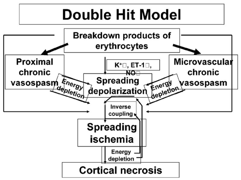
Conclusion: A key issue is the recognition of events other than arterial narrowing such as early brain injury and cortical spreading depression and of their contribution to overall mortality and morbidity.


Figures

Figure 1
Figure 1
A putative pathway for the production of bilirubin-oxidized fragments (BOXes) from blood present post-SAH. Key steps are the liberation of heme from blood and oxidation from free radicals. The dotted line between the heme and oxidation of bilirubin is a pathway for the production of BOXes that has not yet been demonstrated
Figure 2
Figure 2
Relations among Rho kinase, Rho A, PKCI′ and PKCI± for the regulation of myosin and actin in the contraction and relaxation of vascular smooth muscle cells. Rho kinase and Rho A activate myosin light chain (MLC) phosphorylation directly. Rho kinase and Rho A inhibit MLC phosphatase, resulting in long lasting MLC phosphorylation. They also activate PKCI′, which enhances the contraction of vascular smooth muscle cells. PKCI± independently activates the contraction of vascular smooth muscle cells. Caldesmon is an actin-side regulatory protein acting on the detaching between actin and MLC and relaxation of the vascular smooth muscle cells. PKCI′ inhibits the activity of caldesmon through phosphorylates of caldesmon, which causes long lasting interaction between actin and MLC. MLC, myosin light chain; MLCK, myosin light chain kinase; P, phosphorylation; PKC, protein kinase C
Figure 3
Figure 3
Signal transduction for nitric oxide (NO) and endothelin 1 (ET-1). These are antagonistic regulators of cerebral blood flow, released from endothelial cells in response to changes in the shear stress, transmural pressure, concentration of CO2 and O2, ischemia or presence of hemoglobin. NO (vasodilator) and ET-1 (vasoconstrictor) regulate blood vessel tension via smooth muscle cells. NO, due to its high affinity to the heme moiety (1000 times higher than oxygen), stimulates guany(ly)l cyclase, leading to an increase of 3,5′ cyclic guanosine monophosphate and dephosphorylation of MLCs, smooth muscle cell hyperpolarization and closure of calcium channels resulting in vasodilation and an increase of blood flow. ET-1 is a product of several post-translational modifications of pre-pro-ET-1 and big ET-1. It acts on smooth muscles via two types of receptors: ETA, present mostly on smooth muscle cells, whose stimulation leads to smooth muscle constriction (paracrine action), and ETB, present mostly on endothelial cells, stimulation of which leads to an increased NO release and to smooth muscle relaxation (endocrine action). ET-1 stimulation of the ETA receptor leads to the formation of diacylglycerol and inositol 1,4,5-triphosphate, which in turn increases the concentration of intracellular calcium directly or via protein kinase C, resulting in vasoconstriction and decrease of blood flow
Figure 4
Figure 4
Potential mechanisms of increased vascular smooth muscle intracellular Ca2+ and enhanced contraction of cerebral artery myocytes following SAH. Enhanced Ca2+ influx through voltage-dependent Ca2+ channels (VDCCs) may result from a combination of increased VDCC expression (L- and R-type) and increased VDCC activity due to membrane depolarization. Mechanisms contributing to depolarization include oxyhemoglobin-induced internalization of voltage-dependent (KV) K+ channels and decreased activity of large-conductance Ca2+ -activated (BK) K+ channels due to inhibition of Ca2+ sparks and/or increased levels of the cytochrome P450 metabolite 20-hydroxyeicosatetraenoic acid
Figure 5
Figure 5
Double-hit model of delayed ischemic neurological deficits after SAH based on Dreier et al.. The two hits on the brain parenchyma consist of acutely triggered microvascular spasm in response to spreading depolarizations, superimposed on chronic vasospasm

References

    1. Clarke E. Apoplexy in the Hippocratic writings. Bull Hist Med. 1963;37:301. - PubMed
    1. Burnett M, Danish S, McKhann G, II, et al. Pathology and pathophysiology of aneurysmal subarachnoid hemorrhage. In: Leroux P, Winn W, Newell D, editors. Management of Cerebral Aneurysms. Philadelphia, PA: Elsevier; 2004. pp. 127–137.
    1. Weir B, Grace M, Hansen J, et al. Time course of vasospasm in man. J Neurosurg. 1978;48:173–178. - PubMed
    1. Kassell N, Torner J, Haley E., Jr The International Cooperative Study on the Timing of Aneurysm Surgery. Part 1: Overall management results. J Neurosurg. 1990;73:18–36. - PubMed
    1. Dorsch N. Therapeutic approaches to vasospasm in subarachnoid hemorrhage. Curr Opin Crit Care. 2002;8:128–133. - PubMed

Publication types

MeSH terms