. 2009 Jun 10;315(10):1706-14.
doi: 10.1016/j.yexcr.2009.03.006. Epub 2009 Mar 17.

Dynamic analysis of apoptosis using cyanine SYTO probes: from classical to microfluidic cytometry

Affiliations

Dynamic analysis of apoptosis using cyanine SYTO probes: from classical to microfluidic cytometry

Donald Wlodkowic et al. Exp Cell Res. .

Abstract

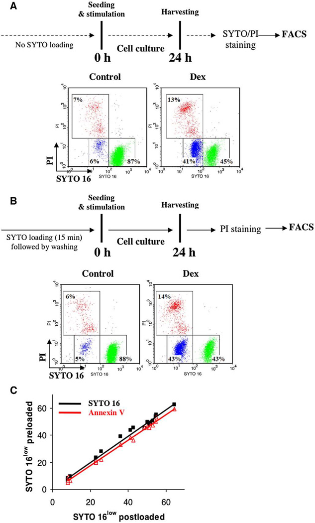
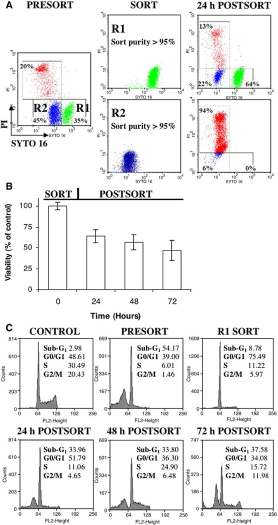
Cell death is a stochastic process, often initiated and/or executed in a multi-pathway/multi-organelle fashion. Therefore, high-throughput single-cell analysis platforms are required to provide detailed characterization of kinetics and mechanisms of cell death in heterogeneous cell populations. However, there is still a largely unmet need for inert fluorescent probes, suitable for prolonged kinetic studies. Here, we compare the use of innovative adaptation of unsymmetrical SYTO dyes for dynamic real-time analysis of apoptosis in conventional as well as microfluidic chip-based systems. We show that cyanine SYTO probes allow non-invasive tracking of intracellular events over extended time. Easy handling and "stain-no wash" protocols open up new opportunities for high-throughput analysis and live-cell sorting. Furthermore, SYTO probes are easily adaptable for detection of cell death using automated microfluidic chip-based cytometry. Overall, the combined use of SYTO probes and state-of-the-art Lab-on-a-Chip platform emerges as a cost effective solution for automated drug screening compared to conventional Annexin V or TUNEL assays. In particular, it should allow for dynamic analysis of samples where low cell number has so far been an obstacle, e.g. primary cancer stems cells or circulating minimal residual tumors.


Figures

Fig. 1
Fig. 1
Intracellular retention of green and red fluorescent SYTO probes: Human B-cell lymphoma cells were pre-stained with 100 nM of SYTO 11/16/62 for 20 min at RT. Following incubation, cells were washed and cultured in dye-free medium for the time indicated. Filled histograms denote unstained control cells. A, B, C, and D denote: 0, 24, 48, 72 h of post-staining culture, respectively. Note that apart from retention heterogeneity all probes were sufficiently retained in live cells after 24 h to allow gating of tagged subpopulation. Results are representatives of four independent experiments. Similar data were obtained on U937 and HL60 cells.
Fig. 2
Fig. 2
Cytotoxicity of substituted cyanine SYTO probes: (A) Influence of SYTO probes on cell viability. Human B-cell lymphoma cells were pre-loaded with 250 nM of selected dyes as described before. Following incubation, cells were washed and cultured in dye-free medium for 48 h. Cell viability was estimated by staining with SYTOX Green (for SYTO 17–62) or SYTOX Red (for SYTO 11 –16) stains. Note that all but SYTO 15 probes do not affect cell viability. Similar results were achieved when cells were continuously challenged with 250 nM of SYTO probes (not shown). Results are representatives of four independent experiments; (B) Influence of SYTO probes on cell cycle progression. Human B-cell lymphoma cells were pre-loaded with selected dyes as described in (A). Cells were then washed and cultured in dye-free medium for 48 h. Cells were subsequently collected and fixed in 70% EtOH for 2 h. Cell cycle analysis was performed using standard PI staining protocol. Note lack of cell cycle disturbances and sub-G1 peak following SYTO pre-loading. Similar results were obtained when cells were continuously cultured in the presence of SYTO dyes (not shown). Results are representatives of four independent experiments; SD values were lower than ±6 for each phase of the cell cycle; (C) Influence of SYTO probes on DNA replication. DNA synthesis (deemed as cell proliferation) was assessed using [methyl-3H]-thymidine incorporation assay 24 h after SYTO staining. Results represent mean (±SD, n=3) of [methyl-3H]-thymidine incorporation relative to untreated controls. Note disturbance of DNA replication for only SYTO 15 stain (p < 0.05). Similar results were achieved when U937 and HL60 cells were continuously cultured in the presence of up to 1 µM of selected SYTO probes (not shown); and (D) The influence of SYTO probes on cell growth. Lymphoma cell lines were treated with indicated SYTO dyes at 250 nM, and the number of viable cells was assessed using standard Trypan Blue assay during the 3-day study. Note cell growth disturbance for SYTO 15 (p<0.05). The results represent mean of at least three independent experiments; normalized SD values were lower than ±3.
Fig. 3
Fig. 3
– Dynamic quantification of apoptosis using SYTO probes: (A) Human B-cell lymphoma cells were exposed to a pro-apoptotic drug dexamethasone (Dex; 1 µM) for 24 h. After stimulation cells were collected and stained according to a standard SYTO 16/PI protocol (end-point assay, upper panel). Data were acquired using FACSCalibur flow cytometer equipped with 488 nm excitation line and standard optical filter configuration. Green events (R1; SYTOhigh) – live cells, Blue events (R2; SYTOlow) – apoptotic cells, Red events (R3; SYTOlow/PI+) – late apoptotic/necrotic cells; (B) As in A) but cells were pre-loaded with 250 nM of SYTO 16, washed, seeded in SYTO-free medium with or without the indicated apoptotic stimuli, and labeled with PI at the end of experiment (dynamic assay, upper panel). Note excellent agreement between results obtained with end-point (A) against dynamic protocols; and (C) Sensitivity of dynamic SYTO assay. Cells were treated with dexamethasone (0–1000 nM) and cycloheximide (0–10 µg/ml) for 6–72 h. After stimulation parallel samples were collected and processed separately according to end-point (SYTO 16 post-loaded) or dynamic (SYTO 16 pre-loaded) protocols. Note comparable sensitivity for both assays over a broad range of stimuli and exposure times (R2≥0.96 for p < 0.05 in Pearson and Lee linear correlation test). For comparison, data from the Annexin V/PI assay were superimposed (red) and indicate exceptional sensitivity of both SYTO 16 assays (R2≥0.97 for p < 0.05). (For interpretation of the references to colour in this figure legend, the reader is referred to the web version of this article.)
Fig. 4
Fig. 4
– Live cell sorting with SYTO 16 probe: (A) Human lymphoma cells were loaded with SYTO 16 (250 nM), treated for 24 h with dexamethasone (Dex; 1 µM) to induce apoptosis and electrostatically sorted based on the following gating strategy: R1 – SYTOhigh/PI (green; deemed viable), and R2 – SYTOlow/PI (blue; deemed early apoptotic). Sorted cells from each gate were seeded in a fresh medium, and analyzed by flow cytometry after 24 h of culture (24 h POSTSORT). Note that SYTO-pre-loading allowed to conveniently track phenotype of cells from R1 subpopulation over time. Dynamic labeling reveled that R1 population underwent apoptosis even after withdrawal of cytotoxic drug (accumulation of SYTOdim/PI early apoptotic and SYTOdim/PI+ secondary necrotic cells); (B) Kinetic viability of B-cell lymphoma cells following sorting procedure described in A). Note that a large number of cells (approx. 48% at 72 h) remained viable after withdrawal of cytotoxic drug. The results represent mean of at least three independent experiments; (C) DNA content analysis on selected subpopulations described in A) and B). Cells were collected at indicated steps and fixed in 70% EtOH for 2 h. Cell cycle analysis was performed using standard PI staining protocol. Note: 1) profound disturbance in cell cycle distribution (G1 cell cycle arrest) in R1 (SYTOhigh/PI; deemed viable) subpopulation; 2) release of G1-arrest with concomitant increase of sub-G1 fraction (deemed apoptotic) after withdrawal of cytotoxic drug. The results represent mean of at least two independent experiments; SD values were lower than±7 for each phase of DNA content. (For interpretation of the references to colour in this figure legend, the reader is referred to the web version of this article.)
Fig. 5
Fig. 5
On-chip detection of apoptosis using SYTO 16 and SYTOX Red probes: (A) U937 cells were cultured in the presence of cycloheximide (CHX) for 24 h followed by an immediate SYTO 16/SYTOX Red staining. Parallel samples were separately analyzed using either conventional flow cytometer (BD FACS Calibur, upper panels) or microfluidic chip-based cytometer (Agilent CellLab Chip, lower panels). Data from chip-based system was converted to FCS 2.0 standard and analyzed using bivariate distribution of SYTO 16 (x-axis) vs. SYTOX Red (y-axis). Note excellent correlation between results from these two technologically dissimilar platforms; (B) Sensitivity of microfluidic cytometry. Cells were treated with cycloheximide (CHX; 0–100 µg/ml), staurosporine (STS; 1 µM). camptothecin (CAM; 1–10 µM) for 1–48 h. After stimulation parallel samples were collected, stained with SYTO 16/SYTOX Red and analyzed separately on a conventional flow cytometer (FACSCalibur) or on-chip microcytometer (Agilent CellLab Chip). Note comparable sensitivity for both analytical platforms (R2≥0.85; p < 0.05 for Pearson and Lee linear correlation test).

References

    1. Wlodkowic D, Skommer J, Darzynkiewicz Z. SYTO probes in cytometry of tumor cell death. Cytometry A. 2008;73(6):496–507. - PubMed
    1. Wlodkowic D, Darzynkiewicz Z. Please do not disturb: destruction of chromatin structure by supravital nucleic acid probes revealed by a novel assay of DNA–histone interaction. Cytometry A. 2008;73A(10):877–879. - PMC - PubMed
    1. Martin RM, Leonhardt H, Cardoso MC. DNA labeling in living cells. Cytometry A. 2005;67A:45–52. - PubMed
    1. Wlodkowic D, Skommer J, Pelkonen J. Towards an understanding of apoptosis detection by SYTO dyes. Cytometry A. 2007;71(2):61–72. - PubMed
    1. Wlodkowic D, Skommer J, Hillier Z. Darzynkiewicz, Multiparameter detection of apoptosis using red-excitable SYTO probes. Cytometry A. 2008;73(6):563–569. - PubMed

Publication types