. 2009 Jul 17;390(3):414-27.
doi: 10.1016/j.jmb.2009.03.028. Epub 2009 Mar 17.

Nucleotide- and activator-dependent structural and dynamic changes of arp2/3 complex monitored by hydrogen/deuterium exchange and mass spectrometry

Affiliations

Nucleotide- and activator-dependent structural and dynamic changes of arp2/3 complex monitored by hydrogen/deuterium exchange and mass spectrometry

Wendy D Zencheck et al. J Mol Biol. .

Abstract

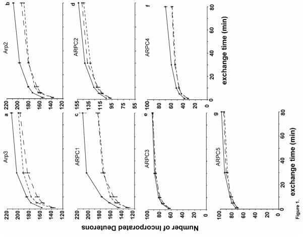
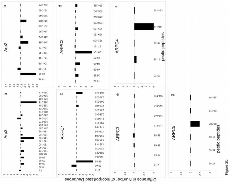
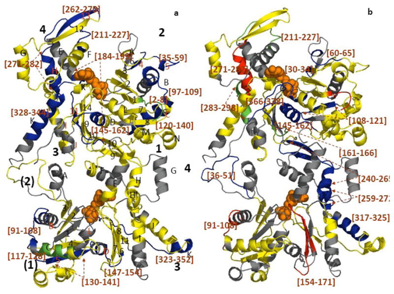
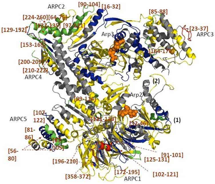
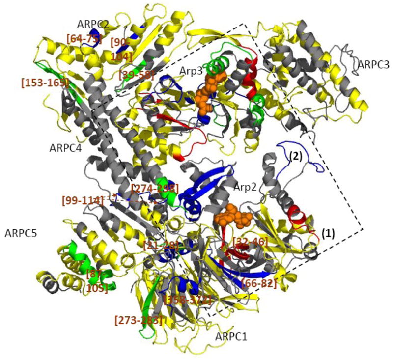
Arp2/3 complex plays a central role in the de novo nucleation of filamentous actin as branches on existing filaments. The complex must bind ATP, protein activators [e.g., Wiskott-Aldrich syndrome protein (WASp)], and the side of an actin filament to form a new actin filament. Amide hydrogen/deuterium exchange coupled with mass spectrometry was used to examine the structural and dynamic properties of the mammalian Arp2/3 complex in the presence of both ATP and the activating peptide segment from WASp. Changes in the rate of hydrogen exchange indicate that ATP binding causes conformational rearrangements of Arp2 and Arp3 that are transmitted allosterically to the Arp complex (ARPC)1, ARPC2, ARPC4, and ARPC5 subunits. These data are consistent with the closure of nucleotide-binding cleft of Arp3 upon ATP binding, resulting in structural rearrangements that propagate throughout the complex. Binding of the VCA domain of WASp to ATP-Arp2/3 further modulates the rates of hydrogen exchange in these subunits, indicating that a global conformational reorganization is occurring. These effects may include the direct binding of activators to Arp3, Arp2, and ARPC1; alterations in the relative orientations of Arp2 and Arp3; and the long-range transmission of activator-dependent signals to segments proposed to be involved in binding the F-actin mother filament.


Figures

Figure 1
Figure 1. Time course of HDX of apo-Arp2/3 complex (solid line), ATP-Arp2/3 complex (short-dashed line), and ATP/WASp-Arp2/3 complex (long-dashed line)
The number of hydrogens exchanged determined by the mass increase for each individual subunit as a function of exchange time. a, Arp3; b, Arp2; c, ARPC1; d, ARPC2; e, ARPC3; f, ARPC4; g, ARPC5.
Figure 2
Figure 2. The changes in deuterium incorporation in peptic peptides in a) ATP-bound vs. apo-Arp2/3 complex and b) ATP- vs. ATP+WASp-Arp2/3 complex
Peptides from each subunit are plotted against the difference in deuterium incorporation following an 80 minute HDX. Differences in deuterium incorporation were determined by the mass difference in individual peptides between a, the apo- and the ATP-bound complex and b, the ATP- and ATP/WASp-bound where negative numbers indicate enhanced protection when ligand is bound, while positive numbers indicate decreased protection. a, Arp3; b, Arp2; c, ARPC1; d, ARPC2; e, ARPC3; f, ARPC4; g, ARPC5.
Figure 3
Figure 3. Mapping the local HDX changes on a ribbon model of the Arp3-Arp2 dimer
a) compares deuterium exchange into individual peptic peptides with and without ATP; b) compares deuterium exchange into individual peptides of ATP-Arp2/3 complex with and without WASp-VCA.) Differences in deuterium incorporation were determined by the mass difference in individual peptides between a) the apo- and the ATP-bound complex and b) the ATP- and ATP/WASp-bound complex. Subdomains of Arp2 and Arp3 are labeled 1–4. Arp3 (top) is based on the crystal structure (1TYQ) and the Arp2 (bottom) is from the composite structure of 1TYQ with modeled subdomains 1 and 2. α-helices are labeled with capital letters and β-sheets are labeled with numbers in a). Segments of the polypeptide chains cleaved by pepsin are labeled by degree of protection: red >+0.5 Da; yellow, −0.5 Da to 0.5 Da; green, −0.5 to −1 Da; blue <−1 Da; and grey, missing peptides. Peptides with significant changes in deuterium incorporation are labeled (brackets).
Figure 4
Figure 4. Mapping the local HDX changes on the model of the Arp2/3 complex with and without ATP
Differences in deuterium incorporation were determined by the mass difference in individual peptic peptides between the apo- and the ATP-bound complex. The subdomains 1 and 2 of Arp2 are from the composite model. Peptic peptides are colored as described in Figure 3.
Figure 5
Figure 5. Mapping the local HDX changes on the model of the ATP-Arp2/3 complex with and without WASp-VCA
Differences in deuterium incorporation were determined by the mass difference in individual peptic peptides between the ATP- and the ATP/WASp-bound complex. The subdomains 1 and 2 of Arp2 are from the composite model. Peptidic peptides are colored as described in Figure 3 and 4. The square indicates possible WASp binding sites.

References

    1. Millard TH, Sharp SJ, Machesky LM. Signalling to actin assembly via the WASP (Wiskott-Aldrich syndrome protein)-family proteins and the Arp2/3 complex. Biochem J. 2004;380:1–17. - PMC - PubMed
    1. Pollard TD, Earnshaw WC. Cell Biology. Saunders; Philadelphia: 2004. updated edition edit.
    1. Kelleher JF, Atkinson SJ, Pollard TD. Sequences, structural models, and cellular localization of the actin-related proteins Arp2 and Arp3 from Acanthamoeba. J Cell Biol. 1995;131:385–97. - PMC - PubMed
    1. Gournier H, Goley ED, Niederstrasser H, Trinh T, Welch MD. Reconstitution of human Arp2/3 complex reveals critical roles of individual subunits in complex structure and activity. Mol Cell. 2001;8:1041–52. - PubMed
    1. Machesky LM, Gould KL. The Arp2/3 complex: a multifunctional actin organizer. Curr Opin Cell Biol. 1999;11:117–21. - PubMed

Publication types

MeSH terms

Substances

LinkOut - more resources