. 2009 May 28;113(22):5516-25.
doi: 10.1182/blood-2008-11-188458. Epub 2009 Mar 18.

Reciprocal responsiveness to interleukin-12 and interferon-alpha specifies human CD8+ effector versus central memory T-cell fates

Affiliations

Reciprocal responsiveness to interleukin-12 and interferon-alpha specifies human CD8+ effector versus central memory T-cell fates

Hilario J Ramos et al. Blood. .

Abstract

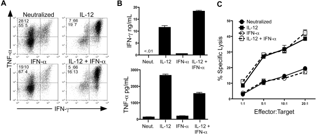
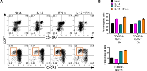
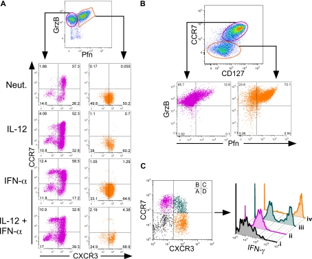
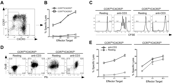
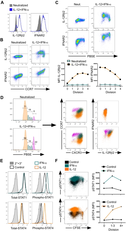
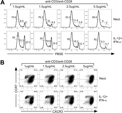
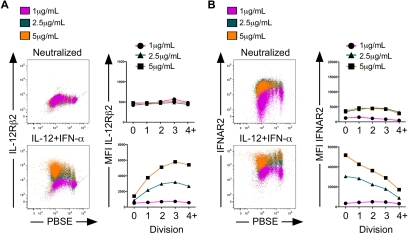
Multiple innate signals regulate the genesis of effector and memory CD8+ T cells. In this study, we demonstrate that the innate cytokines interleukin (IL)-12 and interferon (IFN)-alpha/beta regulate distinct aspects of effector and memory human CD8+ T-cell differentiation. IL-12 exclusively promoted the development of IFN-gamma- and tumor necrosis factor (TNF)-alpha-secreting T effector memory (T(EM)) cells, whereas IFN-alpha drove the development of T central memory (T(CM)) cells. The development of T(EM) and T(CM) was linked to cell division. In rapidly dividing cells, IL-12 programmed T(EM) through induction of the IL-12 receptor beta2. In contrast, IFN-alpha regulated T(CM) development by slowing the progression of cell division in a subpopulation of cells that selectively expressed elevated IFN-alpha/beta receptor-2. The strength of signal delivered through T-cell receptor (TCR) engagement regulated the responsiveness of cells to IL-12 and IFN-alpha. In the presence of both IL-12 and IFN-alpha, these cytokine signals were amplified as the strength of the TCR signal was increased, promoting the simultaneous development of both T(CM) and T(EM). Together, our results support a novel model in which IL-12 and IFN-alpha act in a nonredundant manner to regulate the colinear generation of both effector and memory cells.


Figures

Figure 1
Figure 1
IL-12, but not IFN-α, is sufficient to program human CD8+ T-cell effector functions. (A) Intracellular expression of human IFN-γ and TNF-α from day 7 in vitro polarized human CD8+ T cells. Rested cells were reactivated for 4 hours with PMA and ionomycin in the presence of brefeldin A, and IFN-γ and TNF-α were assessed by intracellular stain and flow cytometric analysis. Data are gated on live, CD8+ cells. (B) Day 7 polarized cells were left unstimulated or stimulated with anti-CD3 for 24 hours, and supernatants were harvested for enzyme-linked immunosorbent assay. Error bars represent the SD of triplicate determinations of each condition. (C) Characterization of CTL activity by 51Cr release assay. Day 7 polarized CD8+ T cells were incubated for 4 hours with 51Cr-labeled THP-1 cells (target) at the E:T ratios shown. CTL activity was assessed by quantification of 51Cr released into the supernatant by β emission. These experiments were performed with 5 different healthy donors with similar results.
Figure 2
Figure 2
Regulation of human CD8+ TCM development by IFN-α. Day 7 cytokine-polarized cells were stained with a panel of anti–human monoclonal antibodies, including CCR7, CD45RA, and CXCR3, to assess memory and effector phenotypes. (A) Analysis of surface markers CCR7 and CD45RA (top panel) and CCR7 and CXCR3 (bottom panel). The induction of CCR7high/CXCR3low cells by IFN-α is indicated by the orange gate. (B) Quantification of human effector and memory profile (top) and chemokine receptor profile (bottom) regulated by IL-12 and IFN-α. Black indicates neutralized; magenta, IL-12; teal, IFN-α; and orange, IL-12 + IFN-α. Error bars represent the SD of triplicate determinations of each condition. These experiments were performed with 7 different healthy donors with similar results.
Figure 3
Figure 3
Human CD8+ TEM and TCM cells display distinct effector properties. Day 7 cytokine-polarized cells were assessed for surface marker and cytokine expression. (A) Cells were gated on live CD8+ cells, and perforin and granzyme B levels were assessed. Cells were gated on either granzyme B single-positive cells (magenta) or perforin–granzyme B double-positive cells (orange) and examined for CCR7 and CXCR3 expression (bottom panels). (B) Live CD8+ cells were gated through either CCR7high, CD127high cells (magenta) or CCR7 and CD127low cells (orange) and assessed for perforin and granzyme B expression (bottom panels). (C) PMA- and ionomycin-activated cells were gated on either CCR7low/CXCR3low (i, black), CCR7high/CXCR3low (ii, magenta), CCR7high/CXCR3high (iii, teal), or CCR7low/CXCR3high (iv, orange), and examined for IFN-γ expression by intracellular staining (right panel).
Figure 4
Figure 4
CCR7 and CXCR3 expression demarcates distinct subpopulations of human CD8+ T cells with functional effector and memory properties. (A) Sorted CD8+ CD45RA+ cells were activated with IL-12 plus IFN-α to day 7. Cells were then sorted into separate CCR7high/CXCR3low or CXCR3high/CCR7low populations. (B) Sorted cells were rested overnight in the absence of IL-2 and subjected to a 51Cr-redirected lysis assay with THP-1 target cells at the indicated E:T ratios. (C) Sorted cells were labeled with CFSE and left untreated (resting) or activated with 1.5 μg/mL plate-bound anti–human CD3 for 3 days (anti-CD3). On day 3, cells were assessed for proliferation by CFSE dilution. (D) Sorted cells were activated, as described in panel C, and examined at day 3 for expression of perforin and granzyme B by bivariant dot plot analysis. (E) Sorted cells were either left untreated (resting) or activated with 1.5 μg/mL anti-CD3 for 3 days (anti-CD3). On day 3, CCR7high/CXCR3low cells (left panel) and CXCR3high/CCR7low cells (right panel) were subjected to a redirected lysis assay, as described above. Each of these experiments was performed twice with 2 separate healthy donors with similar results.
Figure 5
Figure 5
Reciprocal responsiveness to IL-12 and IFN-α/β correlates to development of TEM and TCM cells. (A) Day 3, neutralized or IL-12 plus IFN-α–activated cells were assessed for surface expression of IL-12Rβ2 or IFNAR2 or (B) expression of CCR7, IL-12Rβ2, and IFNAR2. (C) Day 5, PBSE-labeled cells were assessed for IL-12Rβ2 and IFNAR2 expression as a function of division (top), and relative mean fluorescence intensity was quantified (bottom). (D) Day 5, PBSE-labeled cells were gated on division 0 (magenta), division 1-3 (teal), or division 4+ (orange), and chemokine receptor (middle panels) and cytokine receptor (right panels) expression was measured. (E,F) Day 5 cells were polarized with IL-12 + IFN-α, reactivated with cytokines for 30 minutes, and assessed for intracellular STAT or phospho-STAT protein expression. (E) Total STAT, STAT4 (left panels), or phospho-STAT1, phospho-STAT4 (right panels) expression in live CD8+ gated cells. The 2° + 3° antibody alone (gray), unstimulated (black), IFN-α (teal), or IL-12 treated (orange). (F) Dot plot overlays of phospho-STAT1 (top panel, right) or phospho-STAT4 (bottom panel, left) expression as a function of CFSE dilution. Unstimulated (black), IFN-α treated (teal), or IL-12 treated (orange). Quantification of mean fluorescence intensity as a function of CFSE dilution (right panels); phopho-STAT1 (top), phospho-STAT4 (bottom).
Figure 6
Figure 6
TCR signal strength regulates cytokine-dependent TCM development. CD8+ CD45RA+ sorted cells were labeled with PBSE and polarized under either neutralizing or IL-12 + IFN-α conditions with 1 μg/mL, 1.5 μg/mL, 2.5 μg/mL, or 5 μg/mL anti-CD3/anti-CD28 for 5 days. (A) Assessment of division by PBSE dilution as a function of primary activation strength. The percentages of cells that are contained within each gate are indicated above the gate. (B) Cells were analyzed for expression of CXCR3 and CCR7 by bivariant dot plot analysis.
Figure 7
Figure 7
The development of human CD8+ TEM and TCM cells is regulated by both cytokine signaling and strength of primary activation. CD8+ CD45RA+ sorted cells were labeled with PBSE and polarized under neutralizing or IL-12 + IFN-α conditions with 1 μg/mL, 2.5 μg/mL, or 5 μg/mL anti–human CD3 and anti–human CD28 for 5 days. Cells were assessed for expression of IL-12Rβ2 (A) or IFNAR2 (B) as a function of division. Quantification of mean fluorescence intensity is displayed as a function of division for IL-12Rβ2 (A right panels) or IFNAR2 (B right panels) in neutralized (top panels) or IL-12 + IFN-α (bottom panels) cells.

References

    1. Mescher MF, Curtsinger JM, Agarwal P, et al. Signals required for programming effector and memory development by CD8+ T cells. Immunol Rev. 2006;211:81–92. - PubMed
    1. Valenzuela J, Schmidt C, Mescher M. The roles of IL-12 in providing a third signal for clonal expansion of naive CD8 T cells. J Immunol. 2002;169:6842–6849. - PubMed
    1. Curtsinger JM, Valenzuela JO, Agarwal P, Lins D, Mescher MF. Type I IFNs provide a third signal to CD8 T cells to stimulate clonal expansion and differentiation. J Immunol. 2005;174:4465–4469. - PubMed
    1. Casey KA, Mescher MF. IL-21 promotes differentiation of naive CD8 T cells to a unique effector phenotype. J Immunol. 2007;178:7640–7648. - PubMed
    1. Cousens LP, Peterson R, Hsu S, et al. Two roads diverged: interferon α/β- and interleukin 12-mediated pathways in promoting T cell interferon γ responses during viral infection. J Exp Med. 1999;189:1315–1328. - PMC - PubMed

Publication types

MeSH terms