

Window settings for the study of calcified carotid plaques with multidetector CT angiography
- PMID: 19299487
- PMCID: PMC7051539
- DOI: 10.3174/ajnr.A1509
Window settings for the study of calcified carotid plaques with multidetector CT angiography
Abstract
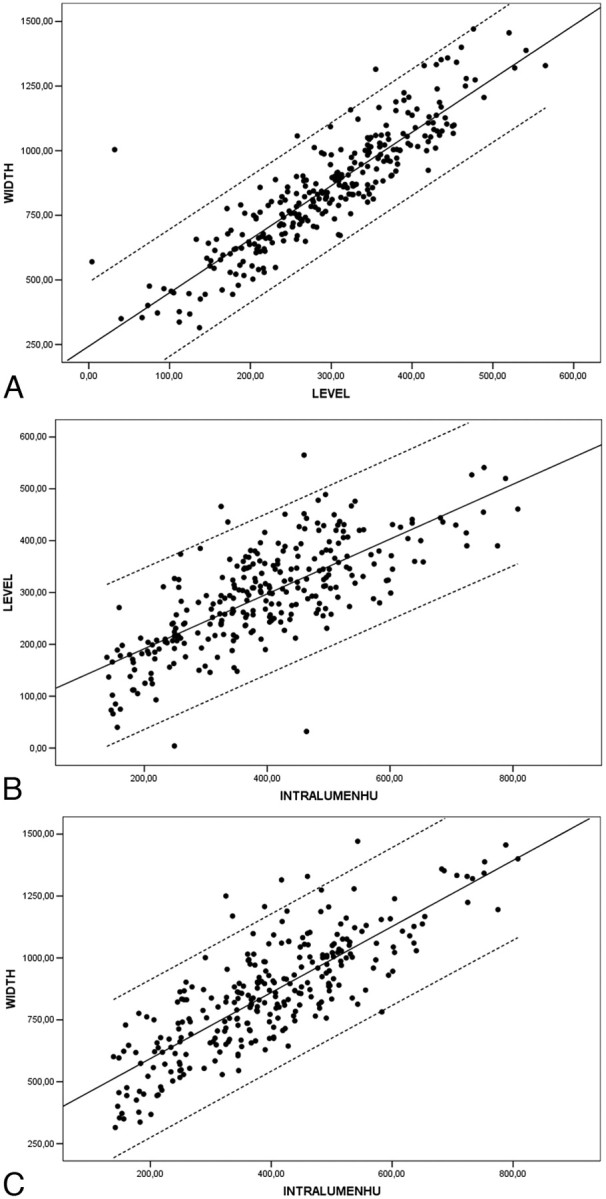
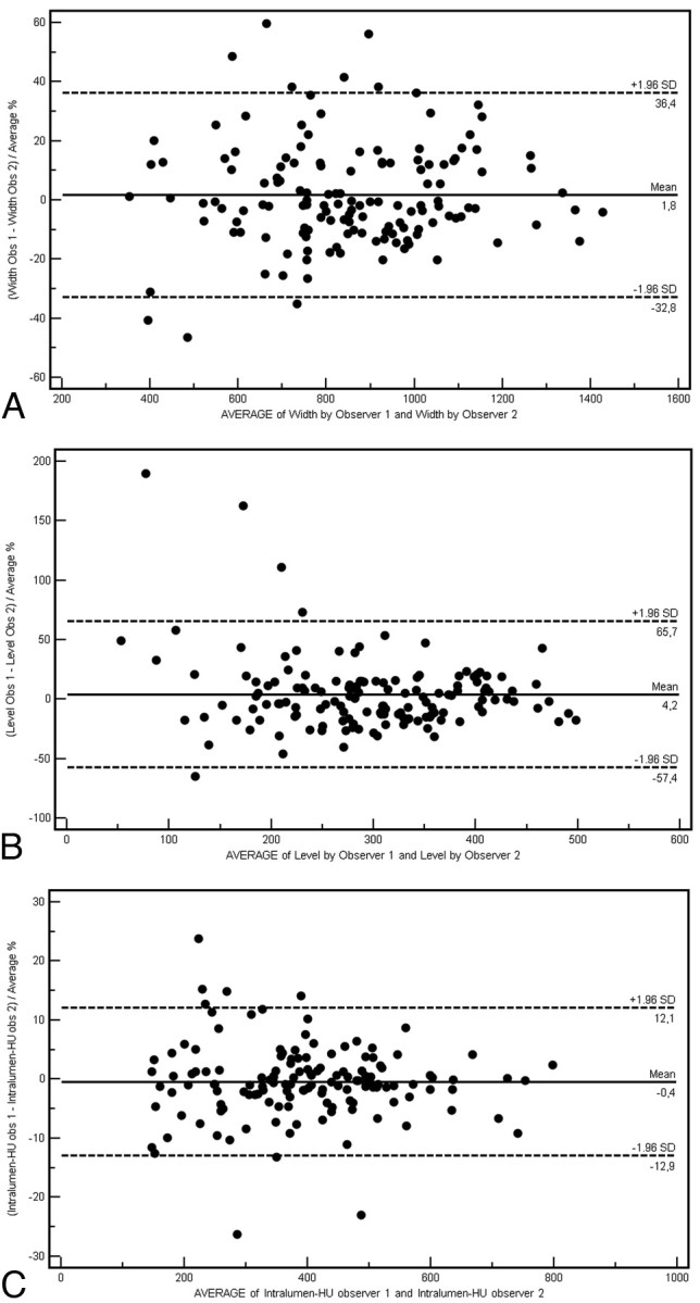
CT angiography (CTA) shows high sensitivity in detecting calcified plaques, but sometimes a bias in the exact quantification of stenosis degree occurs, mainly caused by the high linear attenuation coefficient of the calcified plaques. The purpose of this technical study was to evaluate the most appropriate CT window parameters for the assessment of calcified plaques stating which of them can provide the best inter-observer agreement. Scatter-plots and regression results showed the correlation between both width and level respectively depending on intraluminal Hounsfield units (HU) value (width = intraluminal HU x 2.07; level = intraluminal HU x 0.72). Obtained data indicated that the presence of different stenosis degrees did not modify visualization parameters.
Figures
Similar articles
-
Grading of carotid artery stenosis with multidetector-row CT angiography: visual estimation or caliper measurements?Eur Radiol. 2009 Dec;19(12):2809-18. doi: 10.1007/s00330-009-1508-1. Epub 2009 Jul 18. Eur Radiol. 2009. PMID: 19618190 Free PMC article.
-
Optimizing image contrast display improves quantitative stenosis measurement in heavily calcified coronary arterial segments on coronary CT angiography: A proof-of-concept and comparison to quantitative invasive coronary angiography.Acad Radiol. 2014 Jun;21(6):797-804. doi: 10.1016/j.acra.2014.02.016. Acad Radiol. 2014. PMID: 24809320
-
Performance of semiautomatic assessment of carotid artery stenosis on CT angiography: clarification of differences with manual assessment.AJNR Am J Neuroradiol. 2012 Apr;33(4):747-54. doi: 10.3174/ajnr.A2838. Epub 2011 Dec 22. AJNR Am J Neuroradiol. 2012. PMID: 22194365 Free PMC article.
-
Coronary CTA: stenosis classification and quantification, including automated measures.J Cardiovasc Comput Tomogr. 2009 Nov-Dec;3 Suppl 2:S109-15. doi: 10.1016/j.jcct.2009.10.010. Epub 2009 Oct 30. J Cardiovasc Comput Tomogr. 2009. PMID: 20129518 Review.
-
Measuring progression of coronary atherosclerosis with computed tomography: searching for clarity among shades of gray.J Cardiovasc Comput Tomogr. 2009 Nov-Dec;3 Suppl 2:S81-90. doi: 10.1016/j.jcct.2009.10.011. Epub 2009 Oct 30. J Cardiovasc Comput Tomogr. 2009. PMID: 20129521 Review.
Cited by
-
Evaluating the association between vascular remodeling and plaque calcification patterns of the carotid artery and its effects on ischemic symptoms using CT angiography.Cardiovasc Diagn Ther. 2024 Apr 30;14(2):229-239. doi: 10.21037/cdt-23-428. Epub 2024 Apr 16. Cardiovasc Diagn Ther. 2024. PMID: 38716319 Free PMC article.
-
Assessment of Attenuation in Pericarotid Fat among Patients with Carotid Plaque and Spontaneous Carotid Dissection.AJNR Am J Neuroradiol. 2025 Feb 3;46(2):259-264. doi: 10.3174/ajnr.A8546. AJNR Am J Neuroradiol. 2025. PMID: 39848778
-
Predicting transient ischemic attack risk in patients with mild carotid stenosis using machine learning and CT radiomics.Front Neurol. 2023 Feb 8;14:1105616. doi: 10.3389/fneur.2023.1105616. eCollection 2023. Front Neurol. 2023. PMID: 36846119 Free PMC article.
-
Impact Analysis of Different CT Configurations of Carotid Artery Plaque Calcifications on Cerebrovascular Events.AJNR Am J Neuroradiol. 2022 Feb;43(2):272-279. doi: 10.3174/ajnr.A7401. AJNR Am J Neuroradiol. 2022. PMID: 35121588 Free PMC article.
-
Metal artefact reduction from dental hardware in carotid CT angiography using iterative reconstructions.Eur Radiol. 2013 Oct;23(10):2687-94. doi: 10.1007/s00330-013-2885-z. Epub 2013 May 19. Eur Radiol. 2013. PMID: 23686292
References
-
- Thom T, Haase N, Rosamond W, et al. Heart disease and statistics: 2006 update—a report from the America Heart Association Statistics Committee and Stroke Statistics Subcommitee. Circulation 2006;113:e85–151. Epub 2006 Jan 11 - PubMed
-
- Virmani R, Ladich ER, Burke AP, et al. Histopathology of carotid atherosclerotic disease. Neurosurgery 2006;59:S219–27 - PubMed
-
- Li ZY, Howarth SP, Tang T. Structural analysis and magnetic resonance imaging predict plaque vulnerability: a study comparing symptomatic and asymptomatic individuals. J Vasc Surg 2007;45:768–75 - PubMed
MeSH terms
LinkOut - more resources
Full Text Sources
Medical