. 2009 May 22;284(21):14637-44.
doi: 10.1074/jbc.M807598200. Epub 2009 Mar 19.

Bidirectional signaling through ephrinA2-EphA2 enhances osteoclastogenesis and suppresses osteoblastogenesis

Affiliations

Bidirectional signaling through ephrinA2-EphA2 enhances osteoclastogenesis and suppresses osteoblastogenesis

Naoko Irie et al. J Biol Chem. .

Abstract

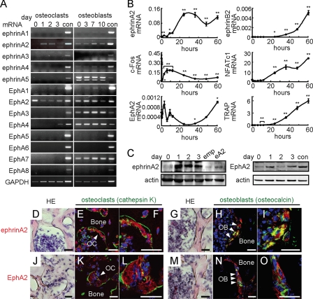
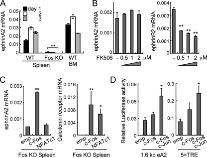
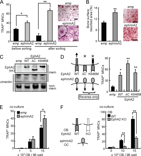
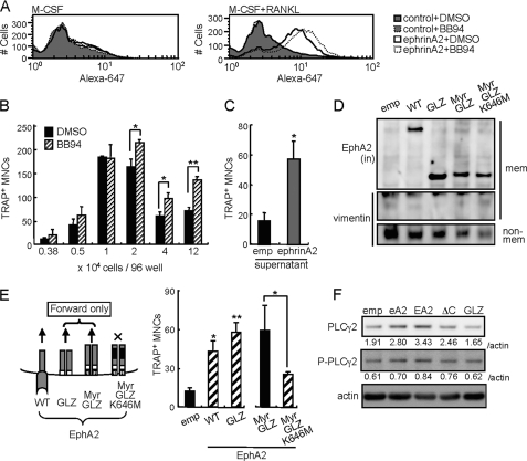
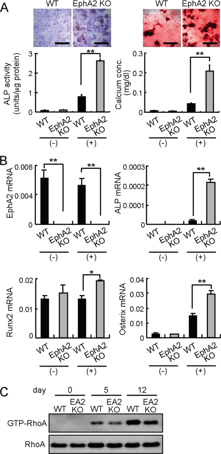
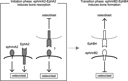
Bone is remodeled constantly throughout life by bone-resorbing osteoclasts and bone-forming osteoblasts. To maintain bone volume and quality, differentiation of osteoclasts and osteoblasts is tightly regulated through communication between and within these two cell lineages. Previously we reported that cell-cell interaction mediated by ephrinB2 ligand on osteoclasts and EphB4 receptor on osteoblasts generates bidirectional anti-osteoclastogenic and pro-osteoblastogenic signals into respective cells and presumably facilitates transition from bone resorption to bone formation. Here we show that bidirectional ephrinA2-EphA2 signaling regulates bone remodeling at the initiation phase. EphrinA2 expression was rapidly induced by receptor activator of NF-kappaB ligand in osteoclast precursors; this was dependent on the transcription factor c-Fos but independent of the c-Fos target gene product NFATc1. Receptor EphA2 was expressed in osteoclast precursors and osteoblasts. Overexpression experiments revealed that both ephrinA2 and EphA2 in osteoclast precursors enhanced differentiation of multinucleated osteoclasts and that phospholipase Cgamma2 may mediate ephrinA2 reverse signaling. Moreover, ephrinA2 on osteoclasts was cleaved by metalloproteinases, and ephrinA2 released in the culture medium enhanced osteoclastogenesis. Interestingly, differentiation of osteoblasts lacking EphA2 was enhanced along with alkaline phosphatase, Runx2, and Osterix expression, indicating that EphA2 on osteoblasts generates anti-osteoblastogenic signals presumably by up-regulating RhoA activity. Therefore, ephrinA2-EphA2 interaction facilitates the initiation phase of bone remodeling by enhancing osteoclast differentiation and suppressing osteoblast differentiation.


Figures

FIGURE 1.
FIGURE 1.
Expression of ephrinA2 and EphA2 during osteoclast and osteoblast differentiation. A, RT-PCR analysis. MDMs were treated with RANKL, and calvarial osteoblasts were treated with ascorbic acid and β-glycerophosphate for the indicated days. con, control adult mouse brain. B, qRT-PCR analysis. MDMs were treated with RANKL for 0, 0.25, 0.5, 1, 3, 6, 9, 12, 24, 36, 48, and 60 h. Error bars represent means ± S.E. (n = 3). *, p < 0.05; **, p < 0.01 versus 0 h. C, immunoblot analysis during osteoclast differentiation. Upper panel, ephrinA2. Negative and positive controls were MDMs infected with empty (emp) and ephrinA2-expressing (eA2) retroviruses, respectively. Lower panel, EphA2. con, control adult mouse brain. day, after RANKL addition. D-O, expression of ephrinA2 and EphA2 in bone. Hematoxylin and eosin (HE) staining of mouse femurs (D, G, J, and M), and immunofluorescence detection of ephrinA2 (red, E, F, H, and I) and EphA2 (red, K, L, N, and O). Osteoclasts (OC) were identified as multinucleated cells expressing cathepsin K (green, E, F, K, and L). Osteoblasts (OB) were cells expressing osteocalcin (green, H, I, N, and O). Higher magnification of osteoclasts or osteoblasts in E, H, K, and N are shown in F, I, L, and O, respectively. Nuclei are shown in blue (4′,6-diamidino-2-phenylindole). Scale bars, 20 μm.
FIGURE 2.
FIGURE 2.
c-Fos-dependent, NFATc1-independent induction of ephrinA2. A, qRT-PCR analysis of ephrinA2 expression. Wild-type (WT) and Fos KO splenocytes and WT bone marrow cells (BM) were cultured in the presence of RANKL for 1-3 days. **, p < 0.01 versus WT spleen control. B, sensitivity of ephrinA2 induction to FK506 at various concentrations (μm). qRT-PCR analysis of ephrinA2 and ephrinB2 was 4 days after RANKL treatment. **, p < 0.01 versus vehicle control (-). C, qRT-PCR analysis of RANKL-induced ephrinA2 (day 1) and calcitonin receptor (day 7) in Fos KO MDMs infected with retroviruses expressing empty cassette (emp), c-Fos, or a constitutively active NFATc1 (NFATc1). *, p < 0.05; **, p < 0.01 versus empty cassette. D, reporter assay using RAW264.7 cells transiently transfected with pGL3 luciferase plasmid driven by the 1.6-kb ephrinA2 promoter (eA2) or the multimerized consensus AP-1-binding sites (5× TRE) and an activator plasmid expressing empty (emp), c-Fos or “single chain” c-Fos∼c-Jun. Transfection efficiency was normalized using Renilla luciferase activity. *, p < 0.05 versus empty cassette. Error bars represent means ± S.E. (n = 3).
FIGURE 3.
FIGURE 3.
EphrinA2 reverse signaling enhances osteoclast differentiation. A, overexpression of ephrinA2 in osteoclast precursors. emp, empty retroviral GFP vector. ephrinA2, ephrinA2-expressing GFP retrovirus. Infected cells (before sorting) and sorted GFP-positive cells (after sorting) were cultured in the presence of M-CSF and RANKL for 3 days. The giant (longitudinal length >125 μm) TRAP+ MNCs were counted. Scale bars, 50 μm. B, bone resorption assay. Infected cells as in A were cultured on bovine bone slices in the presence of M-CSF and RANKL for 10 days. Left panel, values represent bone surface resorbed (%). Right panel, resorption pits (red) are shown. Scale bars, 500 μm. C, immunoblot analysis of EphA2 mutants. MDMs were infected with an empty retrovirus (emp) or viruses expressing EphA2 (WT), EphA2 lacking cytoplasmic region (ΔC), or EphA2 lacking kinase activity (K646M). Membrane (mem) and non-membranous (non-mem) fractions were enriched. EphA2 proteins were detected using anti-EphA2 extracellular domain (ex) antibody. Vimentin was used as a control for non-membranous proteins. D, overexpression of EphA2 mutants in osteoclast precursors. MDMs were infected with empty (emp), EphA2-WT, -ΔC, and -K646M retroviral vectors and were cultured in the presence of M-CSF and RANKL for 4 days. The giant TRAP+ MNCs were counted. E, co-culture of ephrinA2-expressing MDMs with calvarial osteoblasts (OB). MDMs infected with empty (emp) or ephrinA2 retrovirus were seeded at a low cell density (1000 cells/96 wells) together with 1000, 5000, and 15,000 osteoblasts per well. F, osteoclastogenic activity of osteoblasts lacking EphA2. Wild-type MDMs (5000 cells/96 wells) were co-cultured with WT or EphA2-deficient (EphA2 KO) osteoblasts at 5000, 10,000 and 15,000 cells per well. The giant TRAP+ MNCs were counted. Error bars represent means ± S.E. (n = 4-5). *, p < 0.05; **, p < 0.01 versus controls indicated by black bars.
FIGURE 4.
FIGURE 4.
EphA2 forward signaling also enhances osteoclast differentiation. A, fluorescence-activated cell sorter analysis of ephrinA2 on MDMs after 42 h of treatment with either vehicle (DMSO) or MMP inhibitor BB94 in the presence of M-CSF alone (left panel) or M-CSF and RANKL (right panel). control, secondary antibody (Alexa-647) alone. ephrinA2, anti-ephrinA2 and secondary antibodies. B, giant TRAP+ MNCs were counted in cultures of MDMs seeded at various densities and treated with DMSO or BB94 in the presence of M-CSF and RANKL for 4 days. C, effect of conditioned medium of ephrinA2-overexpressing MDMs on osteoclastogenesis. Conditioned medium was prepared by culturing MDMs infected with empty (emp) or ephrinA2 retroviruses for 3 days in the presence of M-CSF and RANKL. Freshly isolated MDMs were cultured in the conditioned medium, and giant TRAP+ MNCs were counted on day 4. D, immunoblot analysis of EphA2 mutants. MDMs were infected with retroviral vectors expressing EphA2 (WT), constitutively active forms of EphA2 lacking the ectodomain (GLZ and Myr-GLZ), or a kinase dead Myr-GLZ (Myr-GLZ-K646M). Membrane (mem) and non-membranous (non-mem) fractions were enriched. EphA2 proteins were detected using anti-EphA2 intracellular (in) domain antibody. Vimentin was used as a control of non-membranous proteins. E, overexpression of EphA2 mutants in osteoclast precursors. MDMs were infected with empty (emp), EphA2-WT, -GLZ, -Myr-GLZ, and -Myr-GLZ-K646M retroviral vectors and were cultured in the presence of M-CSF and RANKL for 4 days. The giant TRAP+ MNCs were counted. F, immunoblot analysis of PLCγ2 and phosphorylated PLCγ2(P-PLCγ2). MDMs were infected with empty (emp), ephrinA2 (eA2), EphA2-WT (EA2), -ΔC, or -GLZ retroviral vectors and were cultured in the presence of M-CSF and RANKL for 2 days. The values indicate intensities of PLCγ2 and P-PLCγ2 bands normalized to actin (relative index). Error bars represent means ± S.E. (n = 3-4). *, p < 0.05; **, p < 0.01 versus controls shown in black bars.
FIGURE 5.
FIGURE 5.
EphA2 signaling inhibits osteoblast differentiation. A, differentiation of wild-type (WT) and EphA2-deficient (EphA2 KO) calvarial osteoblasts. Osteoblast precursors were cultured in the absence (-) or presence (+) of ascorbic acid and β-glycerophosphate. ALP staining (upper left panels) and calcium staining (upper right panels) were performed after 6 and 13 days, respectively. Scale bars, 500 μm. ALP activities and calcium concentrations were quantified. B, qRT-PCR analysis of osteoblast markers in WT and EphA2 KO calvarial osteoblasts cultured under non-osteoblastogenic (-) or osteoblastogenic (+) conditions. RNAs were prepared on day 8. Error bars represent means ± S.E. (n = 3). *, p < 0.05; **, p < 0.01 versus controls shown in black bars. C, RhoA activities in differentiating osteoblasts lacking EphA2. day, after addition of ascorbic acid and β-glycerophosphate.
FIGURE 6.
FIGURE 6.
Schematic presentation of ephrin-Eph interactions during bone remodeling. At the initiation phase of bone remodeling, ephrinA2-EphA2 interaction enhances osteoclastogenesis and inhibits osteoblastogenesis. At the transition phase, ephrinB2-EphB4 interaction inhibits osteoclastogenesis and enhances osteoblastogenesis (11). Note that the effects of ephrin-Eph on osteoclast and osteoblast differentiation are opposite between classes A and B.

References

    1. Karsenty, G., and Wagner, E. F. (2002) Dev. Cell 2 389-406 - PubMed
    1. Teitelbaum, S. L., and Ross, F. P. (2003) Nat. Rev. Genet 4 638-649 - PubMed
    1. Matsuo, K., and Irie, N. (2008) Arch. Biochem. Biophys. 473 201-209 - PubMed
    1. Grigoriadis, A. E., Wang, Z. Q., Cecchini, M. G., Hofstetter, W., Felix, R., Fleisch, H. A., and Wagner, E. F. (1994) Science 266 443-448 - PubMed
    1. Takayanagi, H., Kim, S., Koga, T., Nishina, H., Isshiki, M., Yoshida, H., Saiura, A., Isobe, M., Yokochi, T., Inoue, J., Wagner, E. F., Mak, T. W., Kodama, T., and Taniguchi, T. (2002) Dev. Cell 3 889-901 - PubMed

Publication types