. 2009 May;40(5):1877-85.
doi: 10.1161/STROKEAHA.108.540765. Epub 2009 Mar 19.

Methazolamide and melatonin inhibit mitochondrial cytochrome C release and are neuroprotective in experimental models of ischemic injury

Affiliations

Methazolamide and melatonin inhibit mitochondrial cytochrome C release and are neuroprotective in experimental models of ischemic injury

Xin Wang et al. Stroke. 2009 May.

Abstract

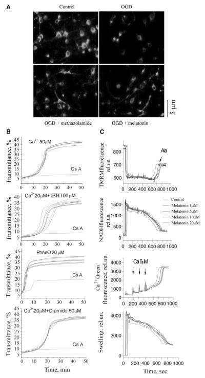
Background and purpose: The identification of a neuroprotective drug for stroke remains elusive. Given that mitochondria play a key role both in maintaining cellular energetic homeostasis and in triggering the activation of cell death pathways, we evaluated the efficacy of newly identified inhibitors of cytochrome c release in hypoxia/ischemia induced cell death. We demonstrate that methazolamide and melatonin are protective in cellular and in vivo models of neuronal hypoxia.

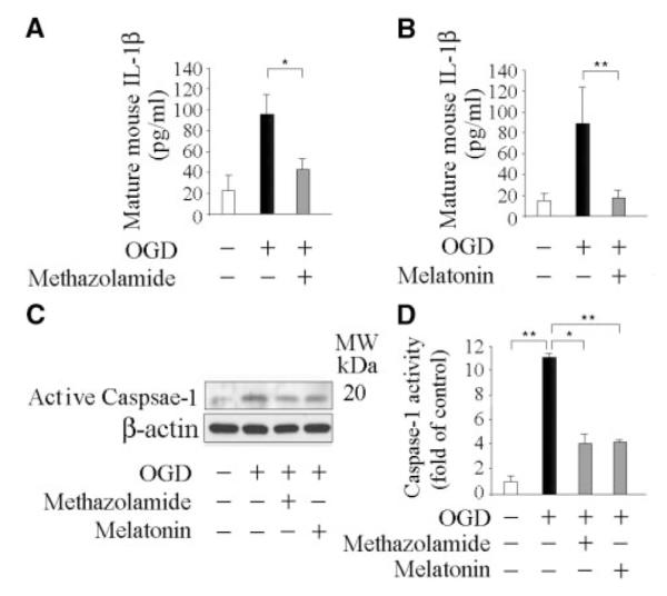
Methods: The effects of methazolamide and melatonin were tested in oxygen/glucose deprivation-induced death of primary cerebrocortical neurons. Mitochondrial membrane potential, release of apoptogenic mitochondrial factors, pro-IL-1beta processing, and activation of caspase -1 and -3 were evaluated. Methazolamide and melatonin were also studied in a middle cerebral artery occlusion mouse model. Infarct volume, neurological function, and biochemical events were examined in the absence or presence of the 2 drugs.

Results: Methazolamide and melatonin inhibit oxygen/glucose deprivation-induced cell death, loss of mitochondrial membrane potential, release of mitochondrial factors, pro-IL-1beta processing, and activation of caspase-1 and -3 in primary cerebrocortical neurons. Furthermore, they decrease infarct size and improve neurological scores after middle cerebral artery occlusion in mice.

Conclusions: We demonstrate that methazolamide and melatonin are neuroprotective against cerebral ischemia and provide evidence of the effectiveness of a mitochondrial-based drug screen in identifying neuroprotective drugs. Given the proven human safety of melatonin and methazolamide, and their ability to cross the blood-brain-barrier, these drugs are attractive as potential novel therapies for ischemic injury.


Figures

Figure 1
Figure 1
Methazolamide and melatonin inhibit cell death of primary cerebrocortical neurons. Cell death of PCNs was induced by 3-hour exposure to OGD with or without a series of concentrations of methazolamide (A and B) and melatonin (C and D). Cell death was evaluated by the lactate dehydrogenase assay (A and C). Data from 3 independent experiments are graphed, and statistically significant differences are indicated with * if P<0.05 and ** if P<0.001. The extent of cell death (as measured by the release of lactate dehydrogenase activity) is always normalized relative to what is measured in the absence of both death stimulus and test drugs (white bar). The black bar corresponds to the extent of cell death in response to the respective stress without the test drug. Gray bars correspond to the extent of cell death with stress and the indicated concentration of the test drug. The relative cell death results are displayed graphically. The resulting curves (plotted semilogarithmically) define the IC50 and maximum protection of methazolamide (B) and melatonin (D).
Figure 2
Figure 2
Methazolamide and melatonin inhibit cell death of primary cerebrocortical neurons under exposure to H2O2 or NMDA. Cell death of PCNs was induced by 18-hour exposure to 1 mmol/L H2O2 (A through C and G) or 500 μmol/L NMDA (D through F and G). A through F, Cell death was evaluated by the lactate dehydrogenase assay. Data from 3 independent experiments are graphed, and statistically significant differences are indicated with * if P<0.05 and ** if P<0.001. White, black, and gray bars are labeled as indicated in Figure 1. G, Phase-contrast light micrographs of control versus H2O2-treated PCNs with or without incubation with methazolamide (10 μmol/L) or melatonin (10 μmol/L) are shown as indicated (middle panel). Nuclei are stained with Hoechst 33342 (upper panel). Arrows point to apoptotic cells with condensed or fragmented chromatin. Bar=5 μm. H, TUNEL staining in cells treated with H2O2 reveals a large increase of positive nuclei with condensed chromatin and DNA fragmentation as compared to control cells. Incubation with methazolamide (10 μmol/L) or melatonin (10 μmol/L) significantly reduces the numbers of TUNEL-positive cells. Scale Bar=5 μm.
Figure 3
Figure 3
Methazolamide and melatonin inhibit the release of mitochondrial apoptogenic factors and forestall caspase-3 activation. Cell death was induced in PCNs by subjecting cells to OGD for 3 hours with or without 10 μmol/L methazolamide (A and C) or 10 μmol/L melatonin (B and C). Subsequently, cells were extracted, and either cytosolic components (A and B) or whole cell lysates (C), or supernatants (D and E) were obtained. The samples, each of which contained 50 μg of protein, were analyzed by Western blot using antibodies to cytochrome c, AIF (cytosolic components; A and B), or caspase-3 (whole cell lysates; C). Beta-actin was used as a loading control. This blot is representative of 3 independent experiments.
Figure 4
Figure 4
Methazolamide and melatonin slow the dissipation of ΔΨm, but melatonin does not inhibit mPT. A, PCNs were subjected to OGD for 3 hours with or without methazolamide (10 μmol/L) and melatonin (10 μmol/L). The living cells were then stained with 2 μmol/L Rh 123 to determine the electrostatic charge of the mitochondria. B, Melatonin was investigated in the in vitro system of purified liver mitochondria that had been stimulated with Ca2+ ions. Melatonin did not prevent the induction of the mPT as judged by an unchanging degree of mitochondrial swelling. The lack of such an effect on addition of melatonin to a solution of stimulated mitochondria is illustrated in B. The same is true of mitochondria stimulated in several ways, ie, with Ca2+ ions; Ca2+ and tBH, PhAsO, or Ca2+ and diamide. The dose—response curves to these stimuli hardly change when melatonin is present at 0.01 μmol/L to 2 mmol/L. In all cases, mitochondrial swelling is monitored by a standard spectroscopic assay using light of 540 nm. C, Liver mitochondria (0.25 mg/mL) were energized with 5 mmol/L glutamate/malate and incubated with 1, 5, 10, and 20 μmol/L of melatonin in buffer containing 250 mmol/L sucrose, 10 mmol/L HEPES, 2 mmol/L KH2PO4. Mitochondria were challenged with bolus additions of 5 μmol/L Ca2+ every 2 minutes until release of sequestered Ca2+ occurred. Alamethicin (100 μg) was added in the end of each sample. Upper left panel is ΔΨm, upper right is in the buffer, lower left is NADH level, and lower right is swelling. See Methods for additional information.
Figure 5
Figure 5
Methazolamide and melatonin inhibit the release of IL-1β and caspase-1 activation. Cell death was induced in PCNs by subjecting cells to OGD for 3 hours with or without 10 μmol/L methazolamide (A, C, and D) and 10 μmol/L melatonin (B, C, and D). Subsequently, conditioned media was collected and assayed for mature IL-1β release after the completion of OGD (A and B), or whole cells were extracted (C and D), and analyzed by Western blot (each of which contained 50 μg of protein) using antibodies to caspase–1 (C). Beta-actin was used as a loading control. This blot is representative of 3 independent experiments. Caspase-1 activity was quantified using a fluorogenic assay in lysed cells. Results are the average of at least 3 independent experiments. *P< 0.05, **P<0.001.
Figure 6
Figure 6
Methazolamide and melatonin diminish damage from cerebral ischemia. Lesion size (A and B) and neuro-scores (C) were determined for mice injected with saline (controls), or methazolamide (20 mg/kg body weight), or melatonin (10 mg/kg body weight). Drugs were administered either 1 hour before or 30 minutes after MCAO. Each test and control group consisted of 7 to 14 mice (n=7 to 14). In all graphs, Data are presented as mean±SE, statistically significant effects are marked with * if P<0.05, and with ** if P<0.001. Brains were quickly removed, cut into coronal sections, and stained with 2,3,5-triphenyltetrazolium chloride (A). In addition, lysates of brain tissue were resolved into cytosolic fractions for analysis by Western blotting (D and E). The protein samples were resolved on SDS/PAGE gels, transferred to nitrocellulose, probed with antibodies to cytochrome c or caspase-3, and reprobed with anti—β-actin. Western blots revealed changes in the cellular localization and degree of maturation of the respective target proteins (D and E). Densitometric scans of these gels allowed the signals from cytoplasmic cytochrome c and activated caspase-3 to be compared to that from β-actin. Each test group consisted of 3 to 5 mice. Again, * indicates that P<0.05 and ** that P<0.001. White bars correspond to brain samples from animals that neither underwent MCAO nor received any test drug. Black bars correspond to samples from saline-injected animals that did undergo MCAO. Grey and light grey bars correspond to samples from test animals, ie, those that were both treated with an experimental drug and underwent MCAO.

References

    1. Li P, Nijhawan D, Budihardjo I, Srinivasula SM, Ahmad M, Alnemri ES, Wang X. Cytochrome c and datp-dependent formation of apaf-1/caspase-9 complex initiates an apoptotic protease cascade. Cell. 1997;91:479–489. - PubMed
    1. Chan PH. Mitochondria and neuronal death/survival signaling pathways in cerebral ischemia. Neurochem Res. 2004;29:1943–1949. - PubMed
    1. Namura S, Nagata I, Takami S, Masayasu H, Kikuchi H. Ebselen reduces cytochrome c release from mitochondria and subsequent DNA fragmen-tation after transient focal cerebral ischemia in mice. Stroke. 2001;32:1906–1911. - PubMed
    1. Zhang WH, Wang X, Narayanan M, Zhang Y, Huo C, Reed JC, Friedlander RM. Fundamental role of the rip2/caspase-1 pathway in hypoxia and ischemia-induced neuronal cell death. Proc Natl Acad Sci U S A. 2003;100:16012–16017. - PMC - PubMed
    1. Wang X, Zhu S, Drozda M, Zhang W, Stavrovskaya IG, Cattaneo E, Ferrante RJ, Kristal BS, Friedlander RM. Minocycline inhibits caspase-independent and -dependent mitochondrial cell death pathways in models of Huntington’s disease. Proc Natl Acad Sci U S A. 2003;100:10483–10487. - PMC - PubMed

Publication types

MeSH terms