Intestinal T cell responses to gluten peptides are largely heterogeneous: implications for a peptide-based therapy in celiac disease
- PMID: 19299713
- PMCID: PMC3306175
- DOI: 10.4049/jimmunol.0803181
Intestinal T cell responses to gluten peptides are largely heterogeneous: implications for a peptide-based therapy in celiac disease
Abstract
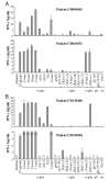
The identification of gluten peptides eliciting intestinal T cell responses is crucial for the design of a peptide-based immunotherapy in celiac disease (CD). To date, several gluten peptides have been identified to be active in CD. In the present study, we investigated the recognition profile of gluten immunogenic peptides in adult HLA-DQ2(+) celiac patients. Polyclonal, gliadin-reactive T cell lines were generated from jejunal mucosa and assayed for both proliferation and IFN-gamma production in response to 21 peptides from wheat glutenins and alpha-, gamma-, and omega-gliadins. A magnitude analysis of the IFN-gamma responses was performed to assess the hierarchy of peptide potency. Remarkably, 12 of the 14 patients recognized a different array of peptides. All alpha-gliadin stimulatory peptides mapped the 57-89 N-terminal region, thus confirming the relevance of the known polyepitope 33-mer, although it was recognized by only 50% of the patients. By contrast, gamma-gliadin peptides were collectively recognized by the great majority (11 of 14, 78%) of CD volunteers. A 17-mer variant of 33-mer, QLQPFPQPQLPYPQPQP, containing only one copy of DQ2-alpha-I and DQ2-alpha-II epitopes, was as potent as 33-mer in stimulating intestinal T cell responses. A peptide from omega-gliadin, QPQQPFPQPQQPFPWQP, although structurally related to the alpha-gliadin 17-mer, is a distinct epitope and was active in 5 out of 14 patients. In conclusion, these results showed that there is a substantial heterogeneity in intestinal T cell responses to gluten and highlighted the relevance of gamma- and omega-gliadin peptides for CD pathogenesis. Our findings indicated that alpha-gliadin (57-73), gamma-gliadin (139-153), and omega-gliadin (102-118) are the most active gluten peptides in DQ2(+) celiac patients.
Figures


Similar articles
-
Gliadin-Specific T-Cells Mobilized in the Peripheral Blood of Coeliac Patients by Short Oral Gluten Challenge: Clinical Applications.Nutrients. 2015 Dec 2;7(12):10020-31. doi: 10.3390/nu7125515. Nutrients. 2015. PMID: 26633487 Free PMC article.
-
T cells in peripheral blood after gluten challenge in coeliac disease.Gut. 2005 Sep;54(9):1217-23. doi: 10.1136/gut.2004.059998. Gut. 2005. PMID: 16099789 Free PMC article.
-
Whole blood interleukin-2 release test to detect and characterize rare circulating gluten-specific T cell responses in coeliac disease.Clin Exp Immunol. 2021 Jun;204(3):321-334. doi: 10.1111/cei.13578. Epub 2021 Feb 28. Clin Exp Immunol. 2021. PMID: 33469922 Free PMC article.
-
Structure of celiac disease-associated HLA-DQ8 and non-associated HLA-DQ9 alleles in complex with two disease-specific epitopes.Int Immunol. 2000 Aug;12(8):1157-66. doi: 10.1093/intimm/12.8.1157. Int Immunol. 2000. PMID: 10917890
-
Coeliac disease--a meeting point for genetics, immunology, and protein chemistry.Lancet. 2003 Apr 12;361(9365):1290-2. doi: 10.1016/s0140-6736(03)12989-3. Lancet. 2003. PMID: 12699968 Review.
Cited by
-
Presence of celiac disease epitopes in modern and old hexaploid wheat varieties: wheat breeding may have contributed to increased prevalence of celiac disease.Theor Appl Genet. 2010 Nov;121(8):1527-39. doi: 10.1007/s00122-010-1408-4. Epub 2010 Jul 28. Theor Appl Genet. 2010. PMID: 20664999 Free PMC article.
-
Genetic and environmental factors affecting the expression of α-gliadin canonical epitopes involved in celiac disease in a wide collection of spelt (Triticum aestivum ssp. spelta) cultivars and landraces.BMC Plant Biol. 2018 Nov 1;18(1):262. doi: 10.1186/s12870-018-1487-y. BMC Plant Biol. 2018. PMID: 30382818 Free PMC article.
-
The Role of Exposomes in the Pathophysiology of Autoimmune Diseases I: Toxic Chemicals and Food.Pathophysiology. 2021 Dec 18;28(4):513-543. doi: 10.3390/pathophysiology28040034. Pathophysiology. 2021. PMID: 35366249 Free PMC article. Review.
-
Gliadin-Specific T-Cells Mobilized in the Peripheral Blood of Coeliac Patients by Short Oral Gluten Challenge: Clinical Applications.Nutrients. 2015 Dec 2;7(12):10020-31. doi: 10.3390/nu7125515. Nutrients. 2015. PMID: 26633487 Free PMC article.
-
The characterization of the repertoire of wheat antigens and peptides involved in the humoral immune responses in patients with gluten sensitivity and Crohn's disease.ISRN Allergy. 2011 Oct 27;2011:950104. doi: 10.5402/2011/950104. Print 2011. ISRN Allergy. 2011. PMID: 23724236 Free PMC article.
References
-
- Jabri B, Sollid LM. Mechanisms of disease: immunopathogenesis of celiac disease. Nat. Clin. Pract. Gastroenterol. Hepatol. 2006;3:516–525. - PubMed
-
- Molberg O, Kett K, Scott H, Thorsby E, Sollid LM, Lundin KE. Gliadin specific, HLA DQ2-restricted T cells are commonly found in small intestinal biopsies from coeliac disease patients, but not from controls. Scand. J. Immunol. 1997;46:103–109. - PubMed
-
- Gianfrani C, Auricchio S, Troncone R. Innate and adaptive immune responses in celiac disease. Immunol. Lett. 2005;99:141–145. - PubMed
Publication types
MeSH terms
Substances
Grants and funding
LinkOut - more resources
Full Text Sources
Other Literature Sources
Medical
Research Materials
Miscellaneous

