. 2009 Apr 1;182(7):4167-74.
doi: 10.4049/jimmunol.0802976.

The cyclic AMP response element modulator {alpha} suppresses CD86 expression and APC function

Affiliations

The cyclic AMP response element modulator {alpha} suppresses CD86 expression and APC function

Martina Ahlmann et al. J Immunol. .

Erratum in

  • J Immunol. 2009 Jul 1;183(1):770

Abstract

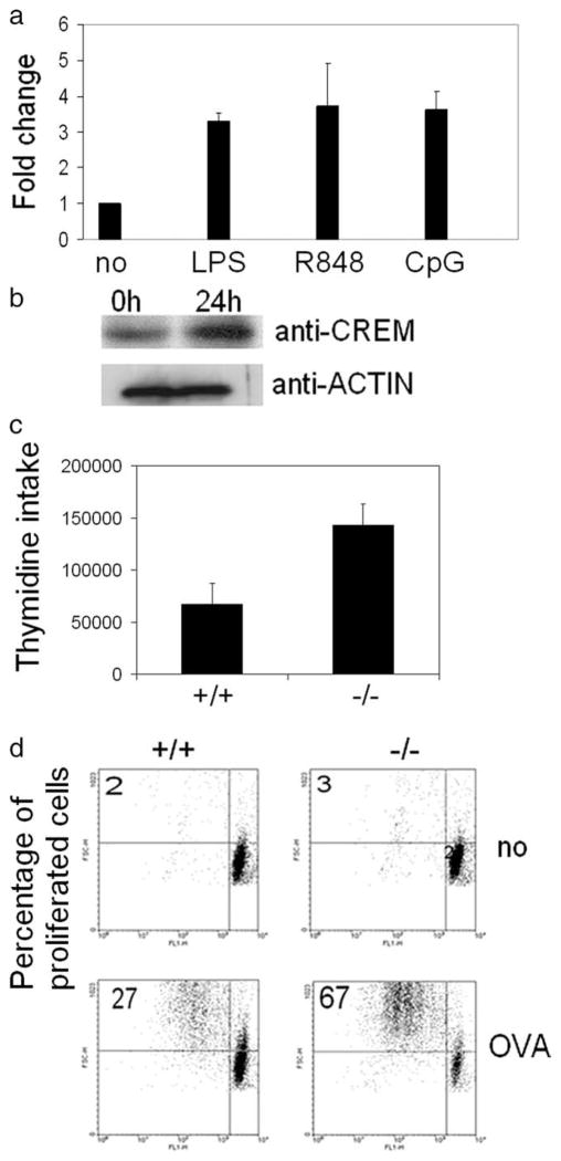
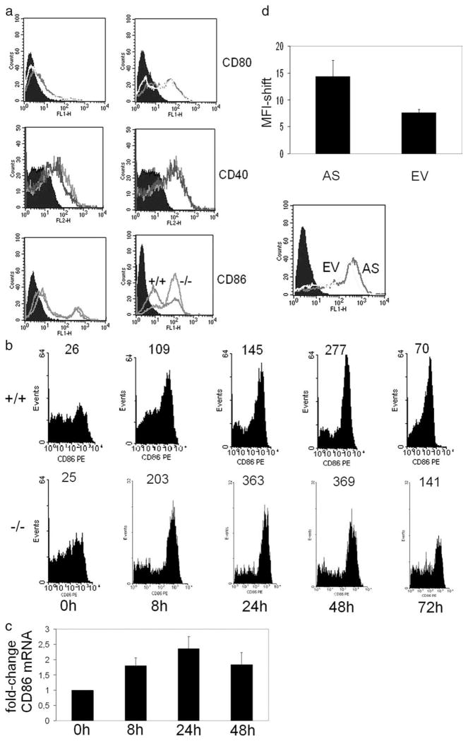
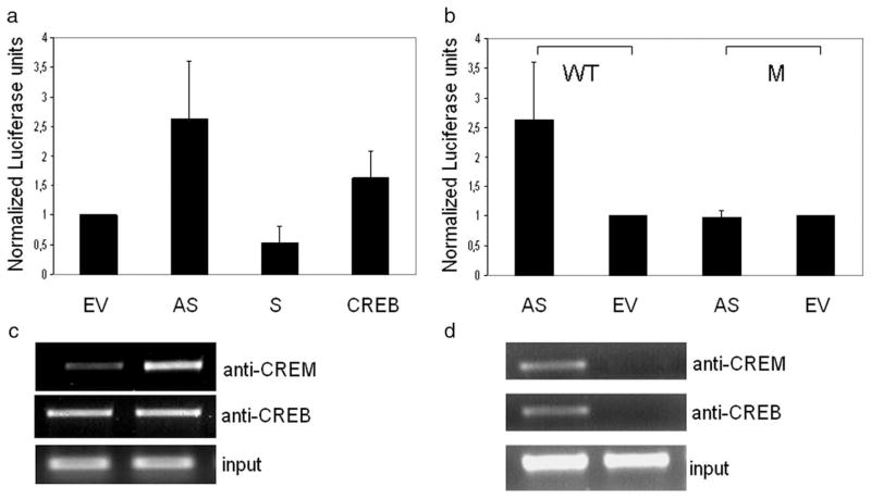
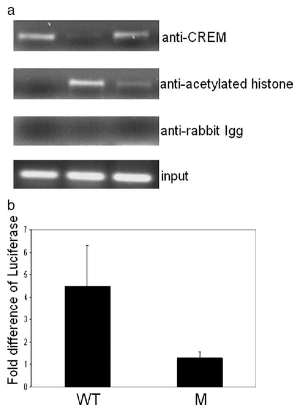
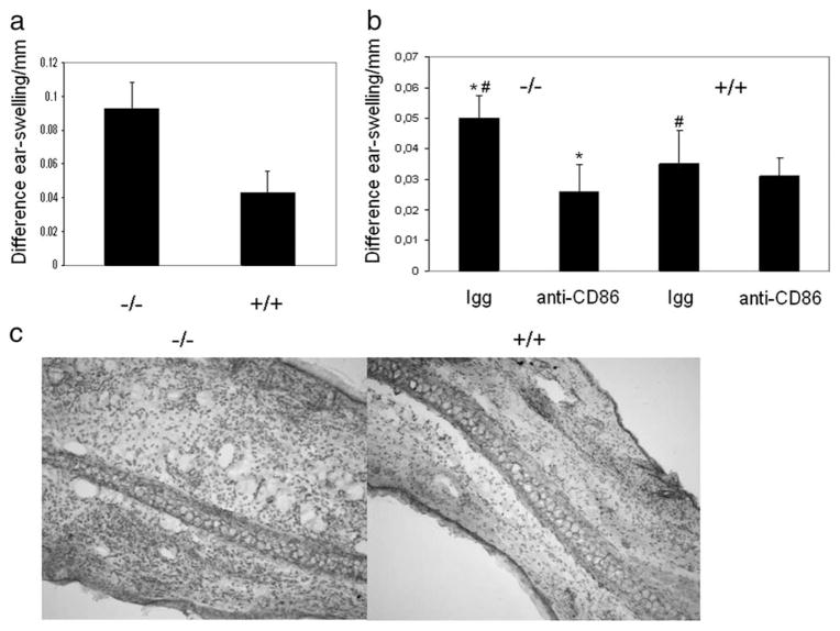
The cAMP response element modulator (CREM)alpha is a widely expressed transcriptional repressor that is important for the termination of the T cell immune response and contributes to the abnormal T cell function in patients with systemic lupus erythematosus. We present evidence that APCs of Crem(-/-) mice express increased amounts of the costimulatory molecule CD86 and induce enhanced Ag-dependent and Ag-independent T cell proliferation. Similarly, human APCs in which CREMalpha was selectively suppressed expressed more CD86 on the surface membrane. CREMalpha was found to bind to the CD86 promoter and suppressed its activity. Transfer of APCs from Crem(-/-) mice into naive mice facilitated a significantly stronger contact dermatitis response compared with mice into which APCs from Crem(+/+) mice had been transferred. We conclude that CREMalpha is an important negative regulator of costimulation and APC-dependent T cell function both in vitro and in vivo.


Conflict of interest statement

Disclosures

The authors have no financial conflict of interest.

Figures

FIGURE 1
FIGURE 1
CREM-deficient DC provide enhanced Ag-specific T cell proliferation. a, BMDCs of C57BL/6 animals were treated with the TLR4 agonist LPS, the TLR7 agonist R848, and the TLR9 agonist CpG for 8 h. RNA was extracted and real-time PCR for CREM was performed and equalized to GAPDH and ribosomal protein L values (n = 3 experiments). b, BMDCs of C57BL/6 animals were stimulated with the TLR4 agonist LPS for 24 h and nuclear proteins were extracted. CREM protein expression was detected by Western blot (n = 2 experiments). c, BMDCs of Crem−/− or Crem+/+ animals were incubated together with T cells from allogenic BALB/c mice. Three days later, T cells were labeled with thymidine and its incorporation was measured 24 h later (n = 5 experiments, p = 0.0065, paired t test). d, BMDCs of DO11.10 Crem−/− or DO11.10 Crem+/+ animals were either left untreated or were pulsed with OVA peptide (1 μM) for 2 h and extensively washed with PBS. BMDCs were cocultured with CFSE-labeled (1 μM) T lymphocytes of OVA-transgenic DO11.10 CREM heterozygote (HZ) mice in ratios ranging from 1:10 to 1:100. FACS analysis of T cells was performed after 3–5 days. Number represents percentage of proliferated cells in each quadrant (n = 4 experiments).
FIGURE 2
FIGURE 2
Enhanced expression of CD86 by CREM-deficient DCs. a, BMDCs from Crem−/− and Crem+/+ animals were either left unstimulated or were stimulated with LPS for 24 h and stained with Abs against CD40, CD80, and CD86. CD86 staining shows the isotypic control (filled histogram), the CD86 expression of the Crem+/+ mice, and the CD86 expression of the Crem−/− mice (n = 3 animals each) for four experiments completed. b, BMDCs from Crem−/− and Crem+/+ animals were either left unstimulated or were stimulated with LPS for depicted time points with Abs against CD86. Number at top of peaks represents mean fluorescent intensity (n = 3 animals each) for two experiments performed. c, BMDCs from Crem+/+ animals were either left unstimulated or were stimulated with LPS for depicted time points. mRNA was extracted and real-time PCR was performed with primers specific for CD86 (n = 3 animals each) for two experiments completed. Data show fold-change relative to gapdh expression. d, U937 cells were transfected with an antisense CREM construct (AS) or an empty-vector construct (EV) together with a GFP construct. CD86 expression on cell surface was measured by flow cytometric analysis 24 h after transfection within a gate set on GFP-positive cells. Mean fluorescence intensity was calculated with error bar representing SEM (n = 6 experiments, p = 0.006, paired t test). Empty vector (gray line histogram) and antisense CREM (black line histogram) are shown (bottom).
FIGURE 3
FIGURE 3
CREM binds to the −21 CRE site of the CD86 promoter. a, The wild-type CD86 promoter luciferase construct has been cotransfected into U937 cells together with an antisense CREMα (AS), a CREMα expression plasmid (S), a CREB expression plasmid or a control plasmid (EV) and cells were stimulated with LPS for 24 h. Luciferase activity was measured and relative luciferase activity was normalized to luciferase activity of the control plasmid. b, The wild-type (WT) or the mutated (M) CD86 luciferase construct was transfected into U937 cells together with either an antisense CREM (AS) plasmid or a control (EV) plasmid and stimulated with LPS. Luciferase activity was measured 24 h after stimulation with LPS (n = 4) and values were normalized to transfection with the control plasmid. c, CD86 promoter luciferase construct has been transfected into U937 cells together with an antisense CREMα (AS) construct or an empty vector (EV) and reporter ChIP has been performed with Abs against CREM and CREB. DNA was amplified with primers specific for the CD86 luciferase construct and run on a 1.5% agarose gel (n = 3). d, CD86 luciferase construct was mutated at the −21 CRE site (M) and transfected into U937 cells. As control, the wild-type luciferase construct (WT) was transfected and an reporter ChIP was performed with Abs against CREM and CREB. DNA was amplified with primers specific for the CD86 luciferase construct and run on a 1.5% agarose gel (n = 3).
FIGURE 4
FIGURE 4
CREM regulates gene expression by a chromatin-dependent mechanism. a, U937 cells were stimulated with LPS for 0, 6, and 24 h and cells were harvested for ChIP analysis with Abs against CREM, acetylated histone 4, and an isotypic control, subsequently semiquantitative PCR with primers specific for the CD86 promoter was performed and the products were run on a 1.5% agarose gel (n = 3 experiments). b, −21 CRE mutated (M) or nonmutated (WT) CD86 promoter luciferase constructs were tranfected into U937 cells and subsequently treated with the HDAC inhibitor SAHA. Luciferase activity after treatment with SAHA was measured 24 h later and values were normalized to nontreated transfected cells. Number shows fold increase of luciferase after treatment with SAHA (n = 3 experiments).
FIGURE 5
FIGURE 5
CREM-deficient DC promote contact dermatitis pathology. a, To haptenize BMDCs in vitro, day 7 BMDCs of Crem−/− and Crem+/+ mice were incubated for 3 h with 1 mM dinitrobenzene sulfonic acid. Cells were extensively washed with PBS and 1 × 10⁶ BMDCs in PBS/mouse were injected i.v. into the tail veins of heterozygous CREM mice on day 0 and mice were challenged on day 6 by painting 10 μl of 0.2% DNFB with an acetone to olive oil ratio of 4:1 on the ventral and dorsal surface of the right ear. The left ear was treated with vehicle alone and served as an internal control for these studies. Contact hypersensitive reaction was determined by the degree of ear swelling measured with a micrometer of the hapten-exposed ear minus that of the vehicle-treated contralateral ear at 24 h after challenge. The y-axis shows difference of ear swelling before and 24 h after challenge with DNFB (n = 5 animals each) in three experiments. Error bar represents SD (p = 0.005, paired t test). b, BMDCs were treated as in a and additionally incubated with an anti-CD86 Ab or an IgG control for 1 h. BMDCs were extensively washed and injected into the tail veins of naive mice. Contact allergy was elicited and measured on day 6 (n = 5 animals each) in two experiments. Error bar represents SD. #, p = 0.028 for isotypic blockade between Crem−/− and Crem+/+ mice.*, p = 0.007 between Crem−/− isotypic control and CD86 blockade. No significant (n.s.) difference was found between Crem+/+ isotypic control and CD86 blockade (by paired t test). c, Animals were sacrificed after 24 h, mouse ears were fixed and stained with H&E. Enhanced cellular infiltration in the ear is shown after transfer of haptenized Crem−/− DCs.

References

    1. Powell JD, Lerner CG, Ewoldt GR, Schwartz RH. The −180 site of the IL-2 promoter is the target of CREB/CREM binding in T cell anergy. J Immunol. 1999;163:6631–6639. - PubMed
    1. Tenbrock K, Juang YT, Tolnay M, Tsokos GC. The cyclic adenosine 5′-monophosphate response element modulator suppresses IL-2 production in stimulated T cells by a chromatin-dependent mechanism. J Immunol. 2003;170:2971–2976. - PubMed
    1. Tenbrock K, Juang YT, Leukert N, Roth J, Tsokos GC. The transcriptional repressor cAMP response element modulator alpha interacts with histone deacetylase 1 to repress promoter activity. J Immunol. 2006;177:6159–6164. - PubMed
    1. Tenbrock K, Juang YT, Gourley MF, Nambiar MP, Tsokos GC. Antisense cyclic adenosine 5′-monophosphate response element modulator up-regulates IL-2 in T cells from patients with systemic lupus erythematosus. J Immunol. 2002;169:4147–4152. - PubMed
    1. Juang YT, Wang Y, Solomou EE, Li Y, Mawrin C, Tenbrock K, Kyttaris VC, Tsokos GC. Systemic lupus erythematosus serum IgG increases CREM binding to the IL-2 promoter and suppresses IL-2 production through CaMKIV. J Clin Invest. 2005;115:996–1005. - PMC - PubMed

Publication types

MeSH terms

Substances