. 2009 Apr 1;182(7):4244-54.
doi: 10.4049/jimmunol.0802041.

Recall responses by helpless memory CD8+ T cells are restricted by the up-regulation of PD-1

Affiliations

Recall responses by helpless memory CD8+ T cells are restricted by the up-regulation of PD-1

Shinichiro Fuse et al. J Immunol. .

Abstract

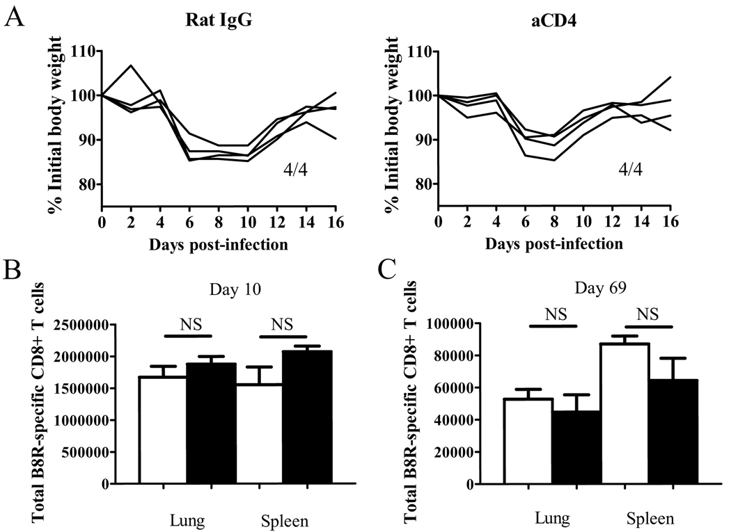
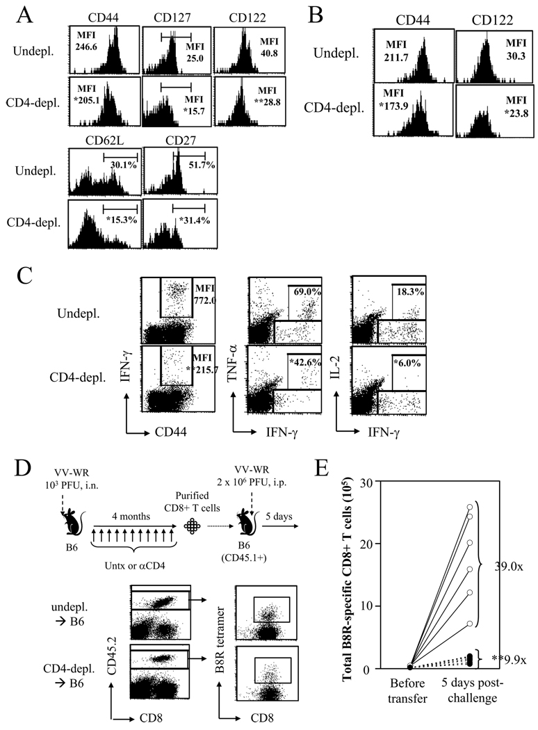
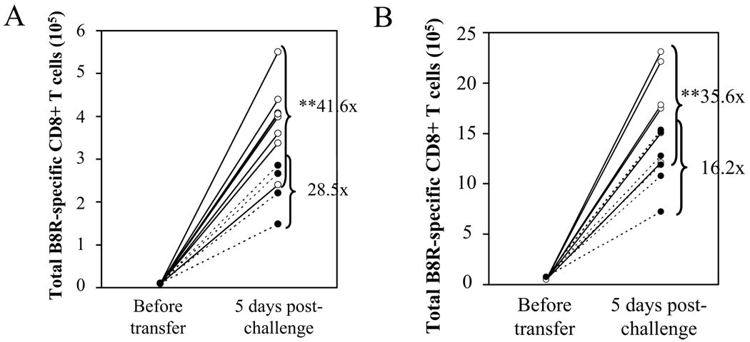
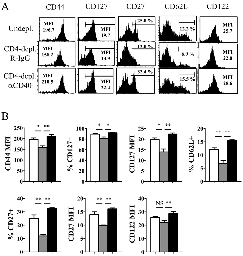
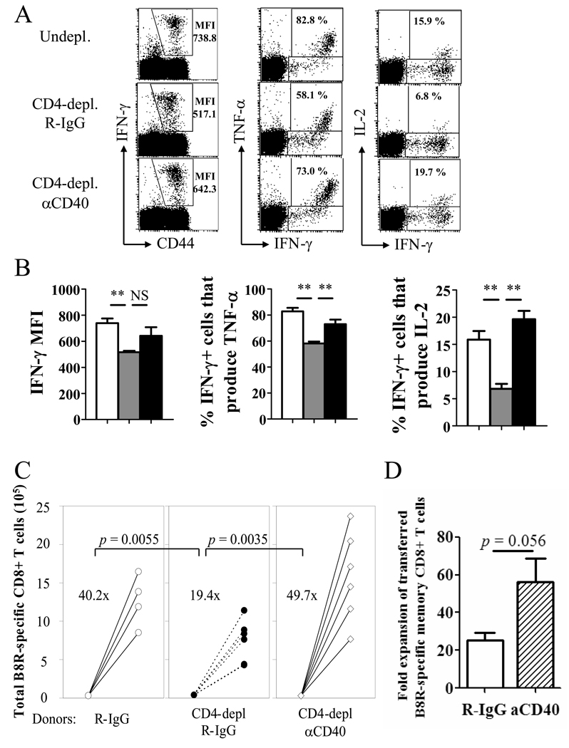
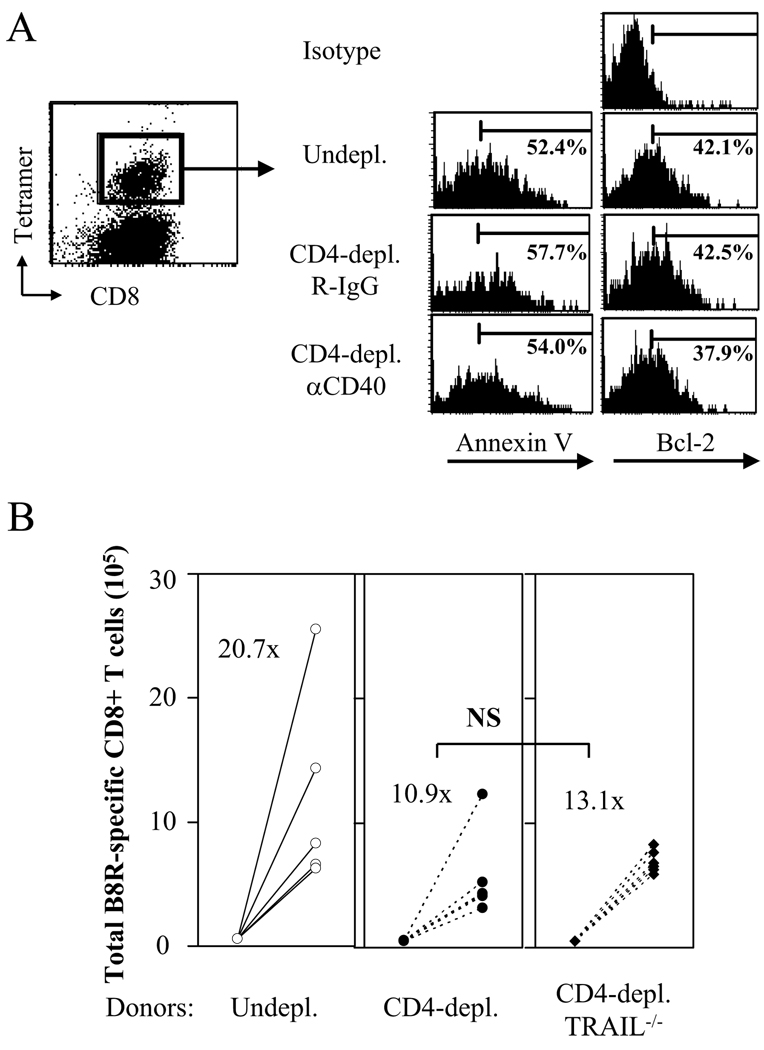
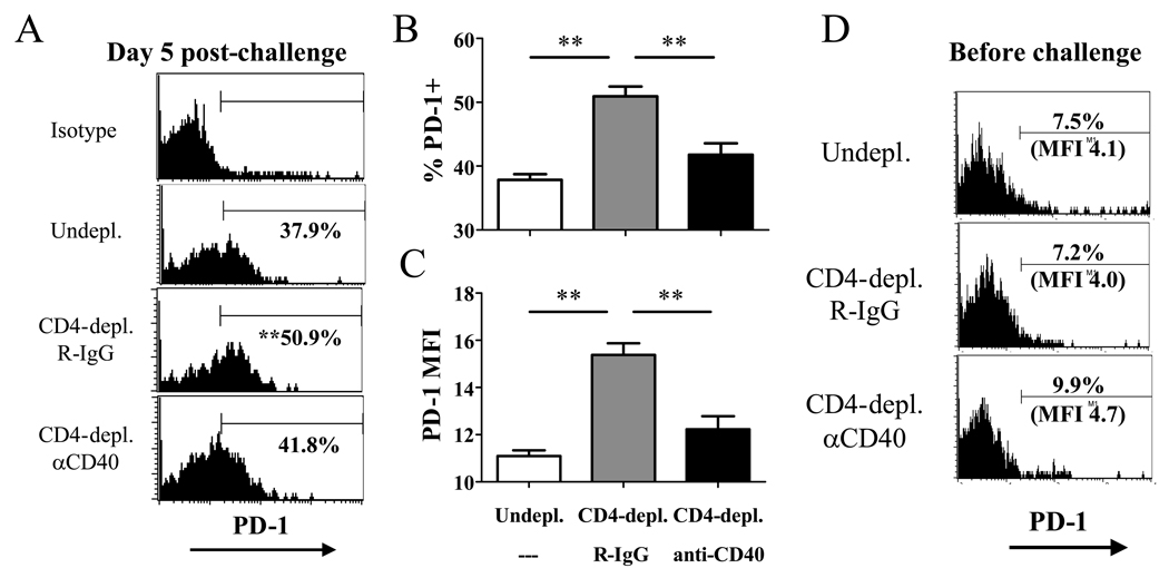
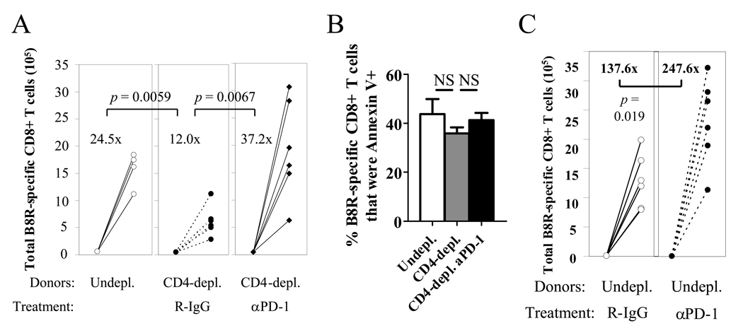
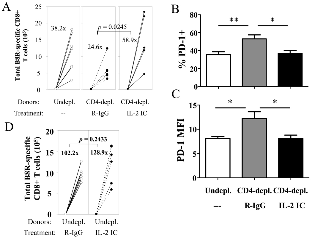
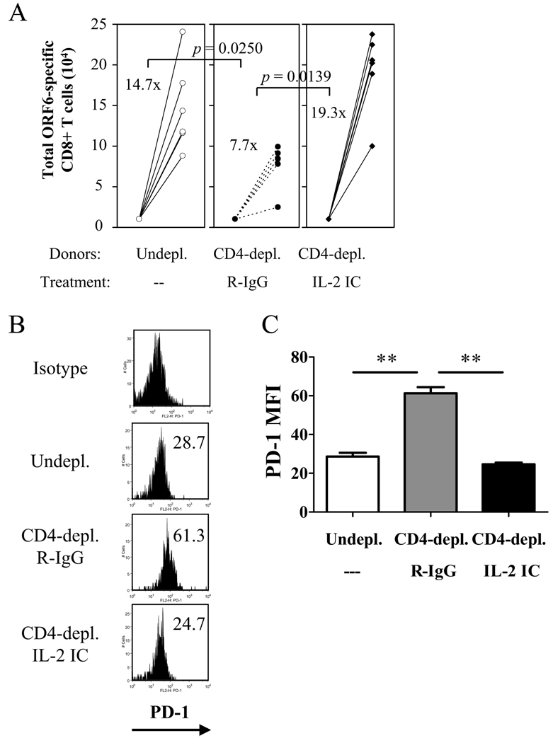
CD4 help is crucial for memory CD8(+) T cell development, yet the mechanisms of CD4 help and why (CD4) helpless memory CD8(+) T cells elicit poor recall responses are currently not well understood. In this study we investigated these questions using an in vivo acute virus infection model. We show herein that CD4 help during priming is required for memory CD8(+) T cell differentiation, and that stimulation of CD40 during priming rescues the helpless defects in the absence of CD4(+) T cells. The defective recall response by helpless memory cells did not correlate with the amount of cell death and was independent of TRAIL. However, helpless memory cells excessively up-regulated the inhibitory receptor PD-1 (programmed cell death-1), and PD-1 blockade enhanced the recall response of helpless memory cells. Furthermore, providing IL-2 signaling in vivo during the recall response reduced PD-1 expression and rescued the recall response of helpless memory cells. Our study identifies molecular pathways involved in CD4 help for memory CD8(+) T cell generation that are independent of TRAIL, and it provides therapeutic implications that helpless memory cell function can be restored at multiple stages through various immunological interventions.


Figures

Figure 1
Figure 1. CD4 help does not affect the initial disease and CD8+ T cell kinetics during VV infection
(A) Weight loss after infection was measured. Numbers indicate surviving mice. (B,C) The VV B8R-specific CD8+ T cell response was measured by tetramer staining on day 10 (B) and day 69 (C) p.i. Total numbers of CD8+ tetramer+ cells in each organ are graphed. White bars: un-depleted, black bars: CD4-depleted. Representative data from two independent experiments with 4 mice per group are shown. Error bars indicate SEM. NS: not significant.
Figure 2
Figure 2. Helpless VV-specific memory CD8+ cells are impaired in function
(A) Phenotype of VV-B8R-specific memory CD8+ cells in un-depleted and CD4-depleted mice were analyzed at day 69 p.i. by tetramer staining. Plots are gated on CD8+ tetramer+ cells. (B) Phenotype of lung-resident memory cells. (C) Cytokine production by VV-specific memory cells was examined by ICCS. Plots are gated on CD8+ cells. Numbers indicate percentage of CD8+ T cells producing IFN-γ (left), or percentage of IFN-γ+ cells that produce TNF-α (middle) or IL-2 (right). (D) The recall response of helped and helpless memory cells were analyzed by an adoptive transfer approach (see Materials and Methods for details). CD8+ T cells were purified from un-depleted or CD4-depleted VV-infected mice 4 months p.i. and were transferred into naïve congenic (CD45.1+) recipients. One day later, recipients were challenged with 2 × 10⁶ PFU of VV-WR, i.p., and 5 days-post challenge, expansion of donor-derived VV-specific memory CD8+ T cells was analyzed by tetramer staining before and after (lower panel) challenge. (E) Results of the adoptive transfer experiment. The total number of donor-derived VV-specific CD8+ T cells analyzed by tetramer staining before transfer and 5 days post-challenge are graphed. Fold expansion of tetramer+ cells was calculated for each recipient, and the number shown indicates the average fold expansion for each group. Each circle (white: undepl. → B6; black: CD4-depl. → B6) represents individual recipients. Representative plots from 2–3 independent experiments, using 3–4 mice per groups are shown. In (D), 4 donor mice per group, and 6–7 recipients mice per group were used. * p > 0.05, ** p > 0.01.
Figure 3
Figure 3. Functional VV-specific memory cells require CD4 help during priming and CD40 signaling
(A) Mice were infected with VV-WR, and were untreated or CD4-depleted during priming (d−1, 0, and 3). 100 days p.i., the recall response of VV-specific memory CD8+ T cells was analyzed by adoptive transfer experiments described in Figure 2. White circles: undepl. → B6; black circles: early CD4-depl. → B6. (B) B6 or CD40−/− mice were infected with VV-WR and the recall response of VV-specific memory CD8+ T cells was analyzed by adoptive transfer and challenge. White circles: B6 → B6; black circles: CD40−/− → B6. Representative data from two independent experiments are shown, using 4 donors per group and 4–8 recipients per group. Each circle represents individual recipients. Fold expansion of tetramer+ cells was calculated for each recipient, and the number shown indicated the average fold expansion for each group. ** p > 0.01.
Figure 4
Figure 4. Providing CD40 stimulation during priming restores phenotypic changes of helpless memory cells
Mice were infected with VV-WR and were un-depleted, CD4-depleted/R-IgG-treated, or CD4-depleted/anti-CD40-treated (d1, 7). (A) VV-specific memory CD8+ T cells were analyzed for expression of phenotypic markers by tetramer staining on day 49 p.i. Plots are gated on CD8+ tetramer+ cells. (B) Graphed presentation of (A). White bars: un-depleted, grey bars: CD4-depleted/R-IgG-treated, black bars: CD4-depleted/anti-CD40-treated. Representative data from two independent experiments with 3–4 mice per group are shown. Error bars indicate SEM. NS: not significant. * p > 0.05, ** p > 0.01.
Figure 5
Figure 5. Providing CD40 stimulation during priming boosts memory cell function in the absence of CD4 help
Mice were infected with VV-WR and were un-depleted, CD4-depleted/R-IgG-treated, or CD4-depleted/anti-CD40-treated (d1, 7). A, Cytokine production by VV-specific memory CD8+ T cells was analyzed by ICCS. Plots are gated on CD8+ cells. Numbers indicate IFN-γ MFI of CD8+ T cells producing IFN-γ (left), or percentage of IFN-γ+ cells that produce TNF-α (middle) or IL-2 (right). B, Graphed representation of (A). White bars: un-depleted, grey bars: CD4-depleted/R-IgG-treated, black bars: CD4-depleted/anti-CD40-treated. Representative data from two independent experiments with 3–4 mice per group are shown. Error bars indicate SEM. NS: not significant. * p > 0.05, ** p > 0.01. C, The recall response by VV-specific memory CD8+ T cells was analyzed by adoptive transfer and challenge. Treatments of donor mice are indicated below. Numbers indicate average fold expansion of VV-specific memory cells for each group. Each circle represents an individual recipient. D, Recall response of helped (undepleted) memory cells that received R-IgG or anti-CD40 during priming is graphed. Fold expansion of transferred (CD45.2+) memory cells is shown (error bars indicate SEM). Representative plots are shown from two independent experiments; consisting of 3–4 mice per group are shown. In C and D, 4 donor mice per group, and 4–6 recipients per group were used.
Figure 6
Figure 6. The defective recall response by helpless memory cells is independent of TRAIL
(A) VV-specific memory CD8+ T cells from un-depleted, CD4-depleted/R-IgG-treated, or CD4-depleted/anti-CD40 treated mice were adoptively transferred into naïve recipients and challenged as shown in Figure 5C. 5 days post-challenge, donor-derived VV-specific CD8+ T cells were stained with Annexin V or by intracellular Bcl-2. Representative plots from two independent experiments with 6 mice per group are shown. Left: representative plot (gated on donor cells) showing gating on tetramer+ cells. Right: gated on CD45.2+ CD8+ tetramer+ cells. (B) The recall response of VV-specific memory CD8+ T cells from un-depleted B6, CD4-depleted B6, or CD4-depleted TRAIL−/− mice was analyzed by adoptive transfer and challenge as shown in Figure 2D. Numbers indicate average fold expansion of VV-specific memory cells for each group. Each circle represents an individual recipient. Representative data from two independent experiments using 4 donor mice per group, 4–6 recipient mice per group are shown.
Figure 7
Figure 7. Helpless memory cells express excess levels of PD-1 during recall compared to helped memory cells
VV-specific memory CD8+ T cells from un-depleted, CD4-depleted/R-IgG-treated, or CD4-depleted/anti-CD40 treated mice were adoptively transferred into naïve recipients and challenged as shown in Figure 5C. 5 days post-challenge, donor-derived VV-specific CD8+ T cells were stained for PD-1 expression. (A) Representative plots gated on CD45.2+ CD8+ tetramer+ cells are shown, and numbers indicate the average percentage of VV-specific CD8+ T cells expressing PD-1. (B) Graphed presentation of (A). (C) MFI of PD-1 expression by donor-derived VV-specific CD8+ T cells. Representative data from two independent experiments with 6 recipients per group are shown. (D) PD-1 expression on resting VV-specific helped and helpless memory CD8+ T cells at 49 days post-infection. Representative data from two independent experiments with 3–4 mice per group are shown. Plots are gated on CD8+ tetramer+ cells. Error bars indicate SEM. * p > 0.05, ** p > 0.01.
Figure 8
Figure 8. Excessive PD-1 expression on helpless memory cells impairs the recall response
(A) Helpless VV-specific memory CD8+ T cells were adoptively transferred and challenged, and were treated with R-IgG or anti-PD-1. 5 days post-challenge, expansion of donor-derived VV-specific CD8+ T cells was analyzed as described in Figure 2D. Numbers indicate average fold expansion of VV-specific memory cells for each group. (B) Donor-derived tetramer+ cells at 5 days post-challenge in the experiment described in (A) were stained with Annexin V. Error bars indicate SEM. (C) Effect of anti-PD-1 on recall response of helped VV-specific memory cells. Representative data from two independent experiments, with 4–6 recipient mice per group are shown. Error bars indicate SEM. * p > 0.05, ** p > 0.01.
Figure 9
Figure 9. IL-2 signaling in vivo rescues the recall response of helpless memory cells
(A) Helpless VV-specific memory CD8+ T cells were adoptively transferred and challenged, and were treated with R-IgG or the IL-2 immune complex. 5 days post-challenge, transferred memory cells were analyzed for expansion as described in Figure 2D. Numbers indicate average fold expansion of VV-specific memory cells for each group. (B,C) Donor-derived tetramer+ cells at 5 days post-challenge were stained for PD-1 expression. Percentage of VV-specific CD8+ T cells expressing PD-1 (B), and MFI of PD-1 expression (C) by donor-derived VV-specific CD8+ T cells is graphed. Error bars indicate SEM. (D) Effect of IL-2 immune complex on recall response of helped VV-specific memory cells. Representative data from two independent experiments, with 4–6 recipient mice per group are shown. * p > 0.05, ** p > 0.01.
Figure 10
Figure 10. PD-1 upregulation by MHV-68-specific helpless memory CD8+ T cells and restoration by IL-2 in vivo
(A) Helpless MHV-68-ORF6-specific memory CD8+ T cells were adoptively transferred and challenged (see Materials and Methods for details), and were treated with R-IgG or the IL-2 immune complex. 5 days post-challenge, transferred memory cells were analyzed for expansion as described in Figure 2D. Numbers indicate average fold expansion of MHV-68-specific memory cells for each group. (B) Donor-derived ORF6 tetramer+ cells at 5 days post-challenge were stained for PD-1 expression. Representative plots gated on CD45.2+ CD8+ cells are shown. Numbers indicate average MFI. (C) MFI of PD-1 expression by donor-derived VV-specific CD8+ T cells is graphed. Representative data from two independent experiments, with 4–6 recipient mice per group are shown. Error bars indicate SEM. ** p > 0.01.

Similar articles

Cited by

References

    1. Kaech SM, Wherry EJ, Ahmed R. Effector and memory T-cell differentiation: implications for vaccine development. Nat Rev Immunol. 2002;2:251–262. - PubMed
    1. Janssen EM, Lemmens EE, Wolfe T, Christen U, von Herrath MG, Schoenberger SP. CD4+ T cells are required for secondary expansion and memory in CD8+ T lymphocytes. Nature. 2003;421:852–856. - PubMed
    1. Shedlock DJ, Shen H. Requirement for CD4 T cell help in generating functional CD8 T cell memory. Science. 2003;300:337–339. - PubMed
    1. Sun JC, Bevan MJ. Defective CD8 T cell memory following acute infection without CD4 T cell help. Science. 2003;300:339–342. - PMC - PubMed
    1. Khanolkar A, Fuller MJ, Zajac AJ. CD4 T cell-dependent CD8 T cell maturation. J Immunol. 2004;172:2834–2844. - PubMed

Publication types

MeSH terms