. 2009 Apr 1;182(7):4395-405.
doi: 10.4049/jimmunol.0803659.

Isoforms of vitamin E have opposing immunoregulatory functions during inflammation by regulating leukocyte recruitment

Affiliations

Isoforms of vitamin E have opposing immunoregulatory functions during inflammation by regulating leukocyte recruitment

Sergejs Berdnikovs et al. J Immunol. .

Erratum in

  • J Immunol. 2010 Jul 15;185(2):1341

Abstract

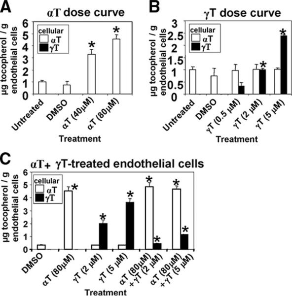
Reports indicate contradictory outcomes for anti-inflammatory functions of the alpha-tocopherol isoform of vitamin E in clinical studies of asthma and atherosclerosis. These seemingly disparate clinical results are consistent with novel unrecognized properties of isoforms of vitamin E reported in this study. We demonstrate that the isoform d-gamma-tocopherol elevates inflammation in experimental asthma. Moreover, d-gamma-tocopherol, at as little as 10% the concentration of d-alpha-tocopherol, ablates the anti-inflammatory benefit of the d-alpha-tocopherol isoform. A mechanism for these opposing immunoregulatory functions of purified tocopherols at physiological concentrations is not through modulation of expression of several cytokines, chemokines, or adhesion molecules, but is, at least in part, by regulation of endothelial cell signals during leukocyte recruitment. These opposing regulatory functions of vitamin E isoforms have impact on interpretations of vitamin E studies. In summary, our studies with purified tocopherol isoforms alter our understanding of vitamin E regulation of vascular function and asthma.


Figures

FIGURE 1
FIGURE 1
Schematic for tocopherol treatments during Ag activation of experimental asthma. A, Structure of natural d-α-tocopherol (d-α-T) and natural d-γ-tocopherol (d-γ-T) that differ by one methyl group (arrows). B, Schematic of time line for tocopherol and OVA treatments. i.n., Intranasal. n = 8–10 animals per group.
FIGURE 2
FIGURE 2
Tocopherol levels and mouse weight on day 21. A, Mouse body weight. B, Wet weight of right lung lobes. C and D, Mice were treated with tocopherols, as in Fig. 1B. Plasma was collected and lungs were perfused free of blood. Plasma or lung tocopherol levels were measured by HPLC. Column A, Mice on chow diet were administered vehicle. Column B, Mice on chow diet were administered d-α-tocopherol (αT). Column C, Mice on chow diet were administered d-γ-tocopherol (γT). Column D, Mice on chow diet were administered both αT and γT. n = 8–10 animals per group. *, p < 0.05 compared with vehicle control.
FIGURE 3
FIGURE 3
Opposite effects of d-α-tocopherol (αT) and d-γ-tocopherol (γT) on leukocyte infiltration into the lung. No effect of αT and γTon blood eosinophil numbers or serum IgE. Mice were treated with tocopherols, as in Fig. 1B. On day 21, the BAL, blood, and lung tissue were examined. A and B, BAL neutrophils, eosinophils, monocytes, and lymphocytes were cytospun and counted by standard morphological criteria. *, <0.05 among groups indicated. **, <0.05 compared with the γT/OVA group. In addition, there is a significant difference between the OVA-treated groups and their corresponding saline groups, with the exception of the lymphocytes in the αT/OVA group, which is not different from the corresponding saline group. C, Representative micrographs of perivascular regions in lung tissue labeled with anti-major basic protein Abs. D, Quantification of perivascular and peribronchial major basic protein+ eosinophils per high powered field (×40 objective). *, p < 0.05 compared with vehicle/OVA-treated mice. In addition, there is a significant difference between the OVA-treated groups and their corresponding saline groups, with the exception of the lymphocytes in the αT/OVA group, which is not different from the corresponding saline group. E, Blood eosinophils. F, Serum OVA-specific IgE was examined by ELISA. *, p < 0.05 compared with saline-treated groups. G, Penh from responses to methacholine using whole body plethysmography on day 21. Arrows are given to assist in comparing the tocopherol-treated, OVA-stimulated groups. Compared with the vehicle, OVA-treated mice (□), the γT/OVA group (arrowhead) elevated the AHR, the αT/OVA group (filled arrow) reduced the AHR, and the α+γT/OVA group (open arrow) had an intermediate phenotype. n = 8–10 animals per group. *, p < 0.05 compared with saline controls. **, The γT groups at 33 and 100 mg/ml methacholine doses were greater than the vehicle/OVA group (p < 0.05).
FIGURE 4
FIGURE 4
The αT and γT treatments did not alter BAL cytokines, tissue eotaxins, lung endothelial cell VCAM-1 expression, or lung tissue PGE2. Mice were treated with tocopherols, as in Fig. 1B. A, B, F, and G, On day 21, the BAL supernatants were examined for cytokines using the CBA Th1/Th2 kit (BD Biosciences). C and D, Lung tissue was placed in RNAlater and then examined by quantitative PCR for eotaxin 1 (CCL11) and eotaxin 2 (CCL24). E, Frozen lung tissue sections were labeled with rat anti-mouse VCAM-1 and a FITC-conjugated secondary Ab. Presented is the sum of fluorescence intensity of the endothelial cells per area of endothelium. Isotype control Ab did not label the tissue (data not shown). H, PGE2 from lung extracts. n = 8–10 animals per group. *, p < 0.05 compared with the corresponding saline control.
FIGURE 5
FIGURE 5
Loading of physiological concentrations of tocopherol in endothelial cells in vitro. Eighty-five percent of confluent monolayers of mHEVa cells were treated overnight with the tocopherols at the concentrations indicated. DMSO at 0.02% is the vehicle control. The cells were washed and weighed, and tocopherol was measured by HPLC/electrochemical detection. A, α-Tocopherol dose curve. B, γ-Tocopherol dose curve. C, Endothelial cells were treated with α-tocopherol and γ-tocopherol individually or together at the indicated concentrations. n = 3–5. *, p < 0.05 compared with vehicle (DMSO) control.
FIGURE 6
FIGURE 6
Tocopherols directly regulate endothelial cells, but not leukocytes, during leukocyte migration without affecting endothelial cell expression of VCAM-1 or MCP-1. A–D, Transwell assay for leukocyte migration with tocopherol left in the culture. Endothelial cells on Transwells were incubated overnight with tocopherols, and then spleen cells were added to the upper chamber of the Transwells to examine cell migration. A, Dose curve of αT. B, Dose curve of γT treatment. C, Dose curve of γT effects on αT (80 μM)-treated cells. D, Endothelial cells were treated overnight with tocopherol, and then in the presence or absence of blocking anti-VCAM-1 Abs, spleen cell migration was determined. Anti-VCAM-1 Abs were added every 4 h, as previously described (19). E and F, Expression of VCAM-1 and MCP-1 by endothelial cells. E, Mean fluorescent intensity of anti-VCAM-1 or isotype Ab control-labeled cells. F, MCP-1 in mHEVa culture supernatant. G–K, Tocopherol pretreatment of cells, followed by washing before the start of the leukocyte migration assay with physiological laminar flow. G, Mice were treated with 2 mg of tocopherol/day or vehicle (ethoxylated castor oil) for 4 days. Spleens were collected, RBC were lysed, and leukocyte tocopherol was determined. H, Spleen leukocytes from untreated mice or spleen leukocytes from mice treated with tocopherols as in G were added to the endothelial cells and examined for transendothelial migration at 15 min under physiological laminar flow. I, Spleen leukocytes from untreated mice or spleen leukocytes from mice treated with tocopherols as in G were added to the endothelial cells and examined for association with the endothelial cells at 2 min under physiological laminar flow. J and K, 85% confluent monolayers of mHEVa cells on slides were treated overnight with the tocopherols at the concentrations indicated to achieve physiological concentrations of tocopherols (Fig. 5), and then the endothelial cells were washed before addition of spleen cells. Spleen cells were isolated from untreated mice, the spleen RBC were lysed, and these spleen leukocytes were added to the endothelial cells to examine leukocyte transendothelial migration under physiological laminar flow. J, Leukocyte transendothelial migration at 15 min under laminar flow. K, Leukocyte association with endothelial cells at 2 min under laminar flow. n = 3–5. *, p < 0.05 compared with DMSO vehicle-treated controls. DMS0 (0.02%) did not affect migration (data not shown).
FIGURE 7
FIGURE 7
Tocopherols regulate VCAM-1 activation of endothelial cell PKCα. Endothelial cells were pretreated overnight with physiological concentrations of tocopherols (80 μM α-tocopherol and/or 2 μM γ-tocopherol), as in Fig. 5. Endothelial cell VCAM-1 was stimulated with anti-VCAM-1 and a secondary Ab for 10 min. Phosphorylation of PKCα-Thr638 was examined by Western blot, developed using an ECL kit, and analyzed by densitometry, as in Materials and Methods. These concentrations of α-tocopherol (80 μM) and γ-tocopherol (2 μM) were optimal for their inhibition or enhancement, respectively, of VCAM-1 activation of PKCα (data not shown). Shown are representative blots and data from five experiments. *, p < 0.05 compared with nonstimulated groups. **, p < 0.05 compared with nonstimulated groups, and p < 0.07 compared with DMSO-treated/anti-VCAM-stimulated group.
FIGURE 8
FIGURE 8
Tocopherol content in cooking oils and human plasma γ-tocopherol levels in several countries. A, Tocopherol in sunflower oil (Presidents Choice), soy oil (Crisco), canola oil (Crisco), corn oil (Mazola), olive oil 1 (Colavita), olive oil 2 (Incecik), olive oil 3 (Phillipo Bario), and olive oil 4 (Eskisigiki) as determined by HPLC/ECD. Shown are mean ± SEM. Bars without error bars indicate errors that are too small to be seen on the graph. B, Human plasma tocopherol levels in several countries (this panel was adapted from review (16)).

References

    1. Abdala-Valencia H, Earwood J, Bansal S, Jansen M, Babcock G, Garvy B, Wills-Karp M, Cook-Mills JM. Nonhematopoietic NADPH oxidase regulation of lung eosinophilia and airway hyperresponsiveness in experimentally induced asthma. Am. J. Physiol. 2007;292:L1111–L1125. - PMC - PubMed
    1. Abdala-Valencia H, Cook-Mills JM. VCAM-1 signals activate endothelial cell protein kinase Cα via oxidation. J. Immunol. 2006;177:6379–6387. - PMC - PubMed
    1. Deem TL, Abdala-Valencia H, Cook-Mills JM. VCAM-1 activation of PTP1B in endothelial cells. J. Immunol. 2007;178:3865–3873. - PMC - PubMed
    1. Deem TL, Cook-Mills JM. Vascular cell adhesion molecule-1 (VCAM-1) activation of endothelial cell matrix metalloproteinases: role of reactive oxygen species. Blood. 2004;104:2385–2393. - PMC - PubMed
    1. Kalayci O, Besler T, Kilinc K, Sekerel BE, Saraclar Y. Serum levels of antioxidant vitamins (α tocopherol, β carotene, and ascorbic acid) in children with bronchial asthma. Turk. J. Peds. 2000;42:17–21. - PubMed

Publication types

MeSH terms