. 2009 Apr 30;41(4):269-76.
doi: 10.3858/emm.2009.41.4.030.

Calcium overload is essential for the acceleration of staurosporine-induced cell death following neuronal differentiation in PC12 cells

Affiliations

Calcium overload is essential for the acceleration of staurosporine-induced cell death following neuronal differentiation in PC12 cells

Su Ryeon Seo et al. Exp Mol Med. .

Abstract

Differentiation of neuronal cells has been shown to accelerate stress-induced cell death, but the underlying mechanisms are not completely understood. Here, we find that early and sustained increase in cytosolic ([Ca2(+)]c) and mitochondrial Ca2(+) levels ([Ca2(+)]m) is essential for the increased sensitivity to staurosporine- induced cell death following neuronal differentiation in PC12 cells. Consistently, pretreatment of differentiated PC12 cells with the intracellular Ca2(+)-chelator EGTA-AM diminished staurosporine-induced PARP cleavage and cell death. Furthermore, Ca2(+) overload and enhanced vulnerability to staurosporine in differentiated cells were prevented by Bcl-XL overexpression. Our data reveal a new regulatory role for differentiation-dependent alteration of Ca2(+) signaling in cell death in response to staurosporine.


Figures

Figure 1
Figure 1
Neuronally differentiated PC12 cells exhibit increased sensitivity to staurosporine-induced cell death. Neuronally differentiated and undifferentiated PC12 cells were treated with indicated concentrations of staurosporine for 12 h (A) or treated with 0.2 µM staurosporine for indicated times (B). The viability of cells was determined by MTT assay. Results are presented as the means ± SD of four independent experiments.
Figure 2
Figure 2
Staurosporine induces DNA fragmentation, caspase-3 activation, and cytochrome c release in neuronally differentiated PC12 cells. Neuronally differentiated and undifferentiated PC12 cells were treated with 0.2 µM staurosporine for the indicated times. (A) The fragmented DNA was analyzed by agarose gel electrophoresis. (B) Total cell extracts were immunoblotted with anti-PARP and anti-β-actin antibodies. (C) Caspase-3 activity was measured using a colorimetric substrate Ac-DEVD-pNA. Values are the means ± SD of three independent experiments. *Significantly different from undifferentiated cells (P < 0.05). (D) Cytoplasmic and mitochondrial fractions of extracts were immunoblotted with anti-cytochrome c antibody. D: differentiated PC12 cells, Un: undifferentiated PC12 cells.
Figure 3
Figure 3
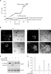
Staurosporine-induced increases in [Ca2+]m and [Ca2+]c are involved in the death of differentiated PC12 cells. (A) Changes in [Ca2+]c were measured in fura-2 loaded neuronally differentiated and undifferentiated PC12 cells using ratiometric fluorescence recording techniques after the application of 0.2 µM staurosporine. In some experiments, cells were pretreated with 20 µM EGTA-AM for 10 min. (B) Changes in [Ca2+]m were monitored in rhod-2 loaded neuronally differentiated (right) and undifferentiated PC12 cells (left), by confocal microscopy, after treatment with 0.2 µM staurosporine for 1 h. (C) Neuronally differentiated PC12 cells were treated with intracellular Ca2+ chelators (20 µM BAPTA-AM or 20 µM EGTA-AM) for 30 min followed by 0.2 µM staurosporine for 3 h. Cell lysates were then immunoblotted with anti-PARP and anti-β-actin antibodies. (D) Neuronally differentiated PC12 cells were treated with 0.2 µM staurosporine for 24 h either in the presence or absence of 10 µM EGTA-AM, and cell viability was measured by MTT assay. Values are the means ± SD of four independent experiments. *Significantly different from cells unexposed to staurosporine (P < 0.05). **Significantly different from cells exposed to staurosporine alone (P < 0.05).
Figure 4
Figure 4
Overexpression of Bcl-XL prevents DNA fragmentation, PARP cleavage, [Ca2+]c increase, and cell death in neuronally differentiated PC12 cells. (A) Bcl-XL overexpressing (Bcl-XL) and control (pCMV) PC12 cells were differentiated and treated with 0.2 µM staurosporine for 24 h, and cell viability was measured by MTT assay. Values are the means ± SD of four independent experiments. *Significantly different from cells unexposed to staurosporine (P < 0.05). **Significantly different from pCMV-transfected cells exposed to staurosporine (P < 0.05). (B) Bcl-XL overexpressing (Bcl-XL) and control (pCMV) PC12 cells were differentiated and treated with 0.2 µM staurosporine for indicated times and the fragmented DNA was analyzed by agarose gel electrophoresis. (C) Bcl-XL overexpressing (Bcl-XL) and control (pCMV) PC12 cells were differentiated and treated with 0.2 µM staurosporine for 6 h, and total cell extracts were immunoblotted with anti-PARP and anti-β-actin antibodies. (D) Changes in [Ca2+]c were measured in fura-2 loaded, neuronally differentiated Bcl-XL overexpressing cells (Bcl-XL) and control (pCMV) using ratiometric fluorescence recording techniques after the application of 0.2 µM staurosporine.

Similar articles

Cited by

References

    1. Beal MF. Mitochondrial dysfunction in neurodegenerative diseases. Biochim Biophys Acta. 1998;1366:211–223. - PubMed
    1. Boise LH, Gonzalez-Garcia M, Postema CE, et al. bcl-x, a bcl-2-related gene that functions as a dominant regulator of apoptotic cell death. Cell. 1993;74:597–608. - PubMed
    1. Demaurex N, Distelhorst C. Cell biology. Apoptosis--the calcium connection. Science. 2003;300:65–67. - PubMed
    1. Desagher S, Martinou JC. Mitochondria as the central control point of apoptosis. Trends Cell Biol. 2000;10:369–377. - PubMed
    1. Dong Z, Saikumar P, Weinberg JM, et al. Calcium in cell injury and death. Annu Rev Pathol. 2006;1:405–434. - PubMed

Publication types

LinkOut - more resources