

The innate immune response and activation of coagulation in alpha1,3-galactosyltransferase gene-knockout xenograft recipients
- PMID: 19300181
- PMCID: PMC4135362
- DOI: 10.1097/TP.0b013e318199c34f
The innate immune response and activation of coagulation in alpha1,3-galactosyltransferase gene-knockout xenograft recipients
Abstract
Background: The role of the innate immune system in the development of thrombotic microangiopathy (TM) after alpha1,3-galactosyltransferase gene-knockout (GTKO) pig organ transplantation in primates is uncertain.
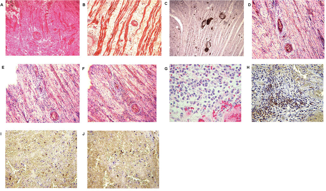
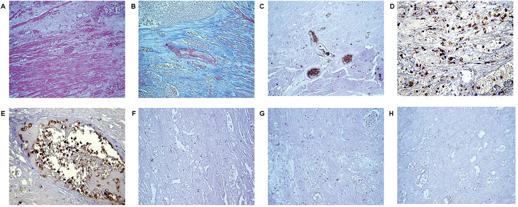
Methods: Twelve organs (nine hearts, three kidneys) from GTKO pigs were transplanted into baboons that received no immunosuppressive therapy, partial regimens, or a full regimen based on costimulation blockade. After graft failure, histologic and immunohistologic examinations were carried out.
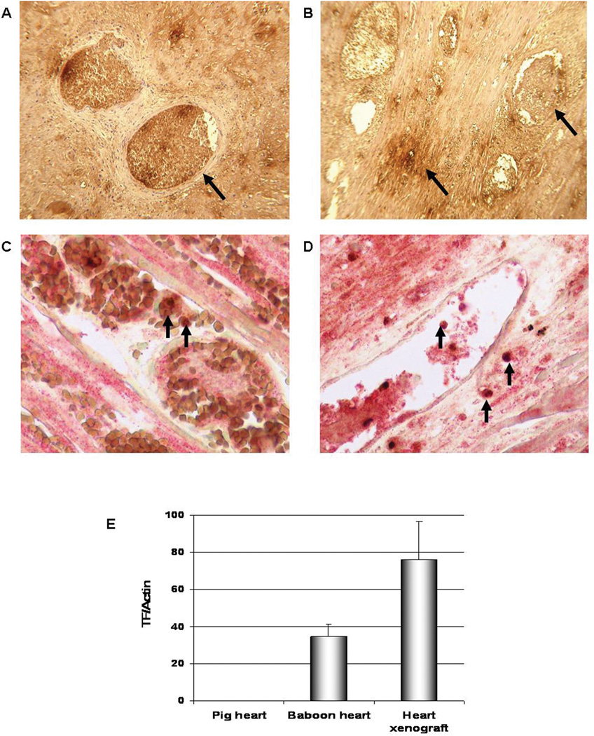
Results: Graft survival of less than 1 day was prolonged to 2 to 12 days with partial regimens (acute humoral xenograft rejection) and to 5 and 8 weeks with the full regimen (TM). Clinical or laboratory features of consumptive coagulopathy occurred in 7 of 12 baboons. Immunohistochemistry demonstrated IgM, IgG, and complement deposition in most cases. Histopathology demonstrated neutrophil and macrophage infiltrates, intravascular fibrin deposition, and platelet aggregation (TM). Grafts showed expression of primate tissue factor (TF), with increased mRNA levels, and TF was also expressed on baboon macrophages/monocytes infiltrating the graft.
Conclusions: Our data suggest that (1) irrespective of the presence or absence of the adaptive immune response, early or late xenograft rejection is associated with activation of the innate immune system; and (2) porcine endothelial cell activation and primate TF expression by recipient innate immune cells may both contribute to the development of TM.
Figures
References
-
- Kuwaki K, Tseng YL, Dor FJ, et al. Heart transplantation in baboons using alpha1,3-galactosyltransferase gene-knockout pigs as donors: initial experience. Nat Med. 2005;11(1):29. - PubMed
-
- Tseng YL, Kuwaki K, Dor FJ, et al. alpha1,3-Galactosyltransferase gene-knockout pig heart transplantation in baboons with survival approaching 6 months. Transplantation. 2005;80(10):1493. - PubMed
-
- Houser SL, Kuwaki K, Knosalla C, et al. Thrombotic microangiopathy and graft arteriopathy in pig hearts following transplantation into baboons. Xenotransplantation. 2004;11(5):416. - PubMed
Publication types
MeSH terms
Substances
Grants and funding
LinkOut - more resources
Full Text Sources
Medical
Miscellaneous
