. 2009 Apr 22;28(8):1099-110.
doi: 10.1038/emboj.2009.62. Epub 2009 Mar 19.

Mimicking Ndc80 phosphorylation triggers spindle assembly checkpoint signalling

Affiliations

Mimicking Ndc80 phosphorylation triggers spindle assembly checkpoint signalling

Stefan Kemmler et al. EMBO J. .

Abstract

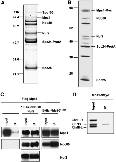
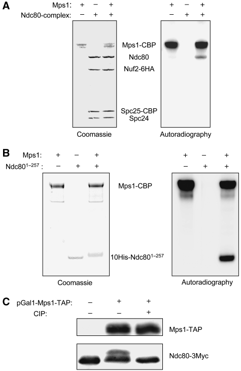
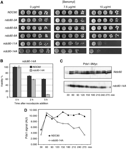
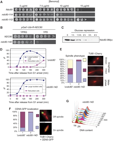
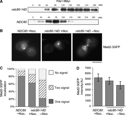
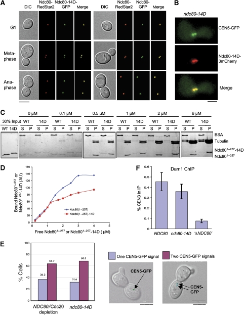
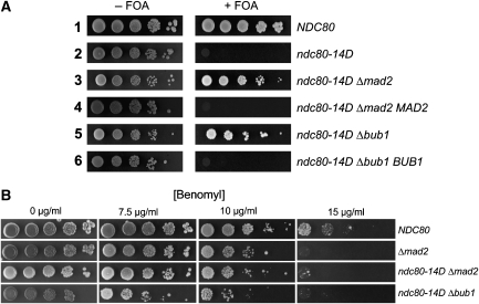
The protein kinase Mps1 is, among others, essential for the spindle assembly checkpoint (SAC). We found that Saccharomyces cerevisiae Mps1 interacts physically with the N-terminal domain of Ndc80 (Ndc80(1-257)), a constituent of the Ndc80 kinetochore complex. Furthermore, Mps1 effectively phosphorylates Ndc80(1-257) in vitro and facilitates Ndc80 phosphorylation in vivo. Mutating 14 of the phosphorylation sites to alanine results in compromised checkpoint signalling upon nocodazole treatment of mutants. Mutating the identical sites to aspartate (to simulate constitutive phosphorylation) causes a metaphase arrest with wild-type-like bipolar kinetochore-microtubule attachment. This arrest is due to a constitutively active SAC and consequently the inviable aspartate mutant can be rescued by disrupting SAC signalling. Therefore, we conclude that a putative Mps1-dependent phosphorylation of Ndc80 is important for SAC activation at kinetochores.


Figures

Figure 1
Figure 1
Mps1 physically interacts with Ndc80 and localizes to the kinetochore. (A) The Ndc80 complex was purified through protein A-tagged Spc24 by affinity chromatography with IgG-sepharose and fractionated by SDS–PAGE. Proteins were identified from Coomassie-stained bands by peptide mass fingerprinting (MALDI-TOF). Bands that are not labelled represent contaminants. (B) As in (A) with the exception that the Ndc80 complex was purified from cells overexpressing Mps1 (strain YSK632). (C) Mps1 and Ndc80 interact in vitro. Flag-Mps1, 10His-Ndc801−257 or 10His-Ndc80 and Nuf2 were co-expressed in E. coli as indicated. 10His-Ndc80 or 10His-Ndc801−257 was affinity purified with NTA-agarose (IP). Input (0.01% of total) and IP (1% of total) were subjected to western analysis with anti-Flag, anti-His and anti-Nuf2 antibodies. (D) ChIP analysis. Mps1-9Myc was immunoprecipitated with anti-Myc antibody. The presence of CEN3 DNA and two flanking DNA regions (ChIII-R and ChIII-L) was analysed by PCR in the input and the immunoprecipitate (IP).
Figure 2
Figure 2
Mps1 phosphorylates Ndc80. (A) In vitro kinase assay. The Ndc80 complex and Mps1 were individually purified from S. cerevisiae by tandem affinity purification and incubated with γ-32P-ATP as indicated. Samples were fractionated by SDS–PAGE and visualized by Coomassie staining plus autoradiography as indicated. CBP, calmodulin-binding peptide. (B) As in (A) with the exception that the His-tagged N-terminal domain of Ndc80 (10His-Ndc801−257) purified from E. coli was used instead of the Ndc80 complex. (C) Mps1 facilitates Ndc80 phosphorylation in vivo. Whole cell extracts were subjected to western analysis with peroxidase-anti-peroxidase (to detect Mps1-TAP) and anti-Myc antibody (to detect Ndc80-3Myc). As shown, control cells (YSK658: NDC80-3MYC) or cells overexpressing Mps1-TAP (YSK658 harbouring plasmid pSK950: pGAL1-MPS1-TAP, 2 μ) were analysed. Extracts were treated with 10 U calf intestinal phosphatase (CIP) for 1 h at 30°C as indicated.
Figure 3
Figure 3
Ndc80 serine and threonine to alanine mutants exhibit a SAC defect. (A) ndc80-11A and ndc80-14A are benomyl sensitive. Serial dilutions of cells with increasing numbers of phosphorylation sites mutated to alanine (see Table II for exact specification of sites mutated) were tested for growth on YPD plates containing the indicated concentration of benomyl. (B) Survival assay. ndc80-14A cells were exposed to nocodazole for the indicated times and then plated onto YPD plates lacking the drug. Viability was calculated by normalizing the number of colonies formed by cells treated with nocodazole to the number of colonies formed by untreated cells (0 h). (C) ndc80-14A fails to maintain long-term Pds1 stability upon nocodazole treatment. α Factor arrested PDS1-9MYC cells were released into medium containing nocodazole. For the indicated time points after the release, Pds1 levels in whole cell extracts were determined by western analysis using anti-Myc antibody. (D) Quantification of (C). AU, arbitrary units.
Figure 4
Figure 4
Phenotype of Ndc80 serine and threonine to aspartate mutants. (A) ndc80-11D is hypertolerant to benomyl. Serial dilutions of cells were tested for growth on YPD plates containing the indicated concentrations of benomyl. (BF) ndc80-14D arrests in metaphase. (B) Serial dilutions of strain YSK1173 (ndc80-14D; PGAL1-Ubi-R-NDC80-9Myc; TUB1-Cherry; CEN5-GFP) and YSK1128 (NDC80; PGAL1-Ubi-R-NDC80-9myc) were tested on YPRG (2% raffinose, 0.1% galactose) or YPD (2% glucose) plates for growth. (C) Ndc80 depletion. Strain YSK1173 was grown to mid-log phase in raffinose medium supplemented with 0.1% galactose. The medium was exchanged for YPD (time point 0), whole cell extracts were derived at the indicated time points and subjected to western analysis with anti-Myc antibody. (D–G) YSK1173 and YSK1155 (PGAL1-Ubi-R-NDC80-9Myc; TUB1-Cherry; CEN5-GFP) (‘Δndc80') were synchronized by α factor treatment in YPD for 3 h and released into YPD medium. (D) Cells with large buds (>2/3 of mother) and cells that exhibited rebudding were counted at the indicated time points (n>100). (E) Spindles were visualized 120 min after the release by fluorescent microscopy (TUB1-Cherry) and quantified as indicated (n>100). Bar: 2 μm. (F) Spindles (TUB1-Cherry) and kinetochores of chromosome V (CEN5-GFP) were visualized 120 min after the release by fluorescent microscopy and quantified as indicated. Bar: 2 μm. (G) The DNA content of YSK1173 was determined by FACS analysis at the indicated time points after the release. α Factor was re-added to the cultures 1 h after the release to arrest cells that executed mitosis in G1 of the next cell cycle. For FACS, data concerning YSK1173 and YSK1155 in the absence of α factor see Supplementary Figure S2.
Figure 5
Figure 5
SAC activation causes the ndc80-14D arrest in metaphase. (AD) Cells were synchronized by α factor treatment and depleted of Ndc80 as in Figure 4D–F. (A) Pds1 levels. At the indicated time points after the release, whole cell extracts of strains YSK1173 (ndc80-14D; PGAL1-Ubi-R-NDC80; PDS1-9MYC) and YSK1128 (NDC80; PGAL1-Ubi-R-NDC80; PDS1-9MYC) were obtained. Pds1 levels were determined by western analysis using anti-Myc antibody. (B) Mad2 localization. Strains YSK1226 (ndc80-14D; PGAL1-Ubi-R-NDC80; MAD2-3GFP) and YMS739 (NDC80; MAD2-3GFP) were analysed by fluorescence microscopy 145 min after the release into YPD with or without nocodazole (Noc.) as indicated. Bar: 5 μm. (C) Quantification of Mad2-3GFP localization. n>100 for each sample. (D) Quantification of Mad2-3GFP signal intensity. The data represent the intensity average of 60 cells (for each sample). When two signals per cell were detected, the values were combined. Error bars represent the standard deviation of the measurements. AU, arbitrary units.
Figure 6
Figure 6
ndc80-14D kinetochores are functional. (A, B) Kinetochore localization of Ndc80-14D. (A) Strain YSK1106 (NDC80-RedStar2; NDC80-GFP) and strain YSK1124 (NDC80-RedStar2; ndc80-14D-GFP) were grown to mid-log phase and analysed by fluorescence microscopy. Bar: 5 μm. (B) Strain YMK1319 (ndc80-14D-Cherry; PGAL1-Ubi-R-NDC80-9Myc; , TetO∷CEN5, TetR-GFP) was depleted of Ndc80 and synchronized by α factor treatment in YPD for 3 h. Cells were analysed 120 min after the release into YPD by fluorescence microscopy. CEN5-GFP marks kinetochore localization. Bar: 2 μm. (C, D) Ndc801−257-14D binds to microtubules in vitro. (C) Taxol-stabilized microtubules at the indicated tubulin dimer concentrations were incubated with purified 1 μM His10-Ndc801−257 (WT) and His10-Ndc801−257-14D (14D) and subsequently centrifuged. About 30% of the pellet (P) and 10% of the supernatant (S) were analysed by SDS–PAGE and Coomassie staining. BSA served as a negative-binding control. (D) His10-Ndc801−257 and His10-Ndc801−257-14D at the indicated concentrations were incubated with or without taxol-stabilized microtubules at 1 μM Tubulin dimer concentration. After centrifugation, 30% of the pellets were subjected to SDS–PAGE plus Coomassie staining and the His10-Ndc801−257 or His10-Ndc801−257-14D bands were quantified. The values for bound His10-Ndc801−257 or His10-Ndc801−257-14D shown in the graph represent the pelleted amount in the presence of microtubules minus the pelleted amount in the absence of microtubules. AU, arbitrary units. (E) ndc80-14D cells exhibit wild-type-like bipolar attachment. Strains YMS412 (NDC80, pGAL-CDC20, TetO∷CEN5, TetR-GFP) and YSK1173 (ndc80-14D, pGAL-Ubi-R-NDC80, TetO∷CEN5, TetR-GFP) were synchronized as in (B). CEN5-GFP was visualized by fluorescence microscopy 120 min after the release. The percentage of cells with one or two CEN5-GFP signals (as shown by DIC/GFP overlays) was quantified (n>100). Bar: 5 μm. (F) Dam1 localization at ndc80-14D kinetochores. Strain YSK1170 (ndc80-14D; PGAL1-Ubi-R-NDC80-9myc), strain YMS412 (NDC80, PGAL1-CDC20) and strain YSK1155 (PGAL1-Ubi-R-NDC80-9Myc) (‘Δndc80') were synchronized by α factor treatment in YPD for 3 h and released into YPD. Cells were subjected to ChIP analysis with anti-Dam1 antibody 120 min after the release. The percentage of CEN3 DNA recovered in the IP is shown in respect to the input. Error bars represent the standard deviation of two independent experiments. Note that under these conditions, YMS412 arrests in metaphase (due to Cdc20 depletion) and, thus, provides the appropriate control for YSK1170 that arrests in metaphase due to the ndc80-14D mutation.
Figure 7
Figure 7
Elimination of SAC signalling rescues ndc80-14D cells. (A) Deletion of MAD2 or BUB1 rescues ndc80-14D mutants. All strains harboured an episomal copy of NDC80 on a CEN/URA3 plasmid. Δmad2 MAD2 or Δbub1 BUB1 cells harboured the wild-type gene copy on a CEN/LEU2 plasmid. All other strains harboured an empty CEN/LEU2 plasmid. Serial dilutions of cells were tested for growth on plates lacking leucine in the absence or presence of FOA. FOA counter selects for the plasmid-based copy of NDC80. (B) Serial dilutions of cells with the specified genotype were tested for growth on YPD plates containing the indicated concentrations of benomyl.

References

    1. Ahonen LJ, Kallio MJ, Daum JR, Bolton M, Manke IA, Yaffe MB, Stukenberg PT, Gorbsky GJ (2005) Polo-like kinase 1 creates the tension-sensing 3F3/2 phosphoepitope and modulates the association of spindle-checkpoint proteins at kinetochores. Curr Biol 15: 1078–1089 - PubMed
    1. Biggins S, Murray AW (2001) The budding yeast protein kinase Ipl1/Aurora allows the absence of tension to activate the spindle checkpoint. Genes Dev 15: 3118–3129 - PMC - PubMed
    1. Castillo AR, Meehl JB, Morgan G, Schutz-Geschwender A, Winey M (2002) The yeast protein kinase Mps1p is required for assembly of the integral spindle pole body component Spc42p. J Cell Biol 156: 453–465 - PMC - PubMed
    1. Chan GK, Liu ST, Yen TJ (2005) Kinetochore structure and function. Trends Cell Biol 15: 589–598 - PubMed
    1. Cheeseman IM, Anderson S, Jwa M, Green EM, Kang J, Yates JR III, Chan CS, Drubin DG, Barnes G (2002) Phospho-regulation of kinetochore-microtubule attachments by the Aurora kinase Ipl1p. Cell 111: 163–172 - PubMed

Publication types

MeSH terms