. 2009 Mar;5(3):e1000334.
doi: 10.1371/journal.ppat.1000334. Epub 2009 Mar 20.

Activation of host translational control pathways by a viral developmental switch

Affiliations

Activation of host translational control pathways by a viral developmental switch

Carolina Arias et al. PLoS Pathog. 2009 Mar.

Abstract

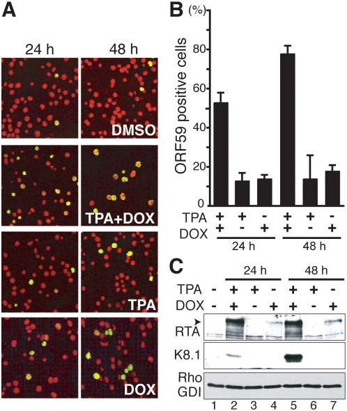
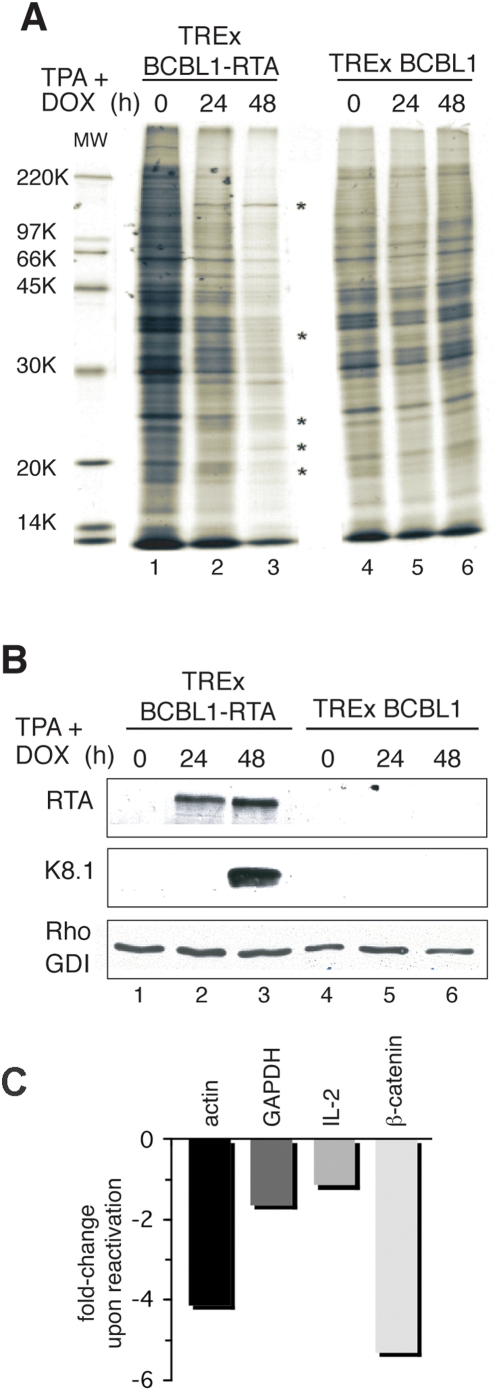
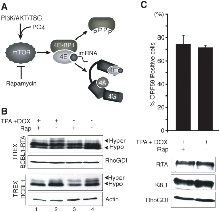
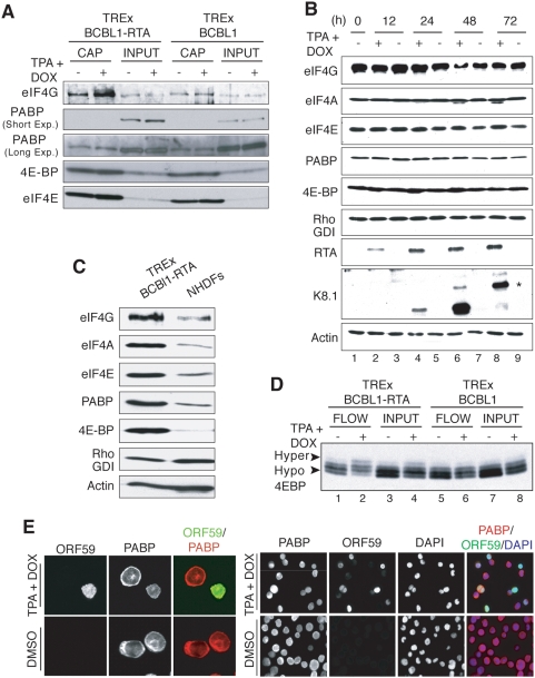
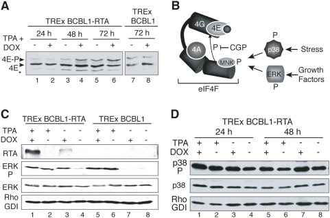
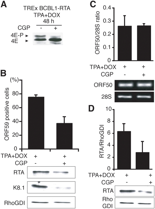
In response to numerous signals, latent herpesvirus genomes abruptly switch their developmental program, aborting stable host-cell colonization in favor of productive viral replication that ultimately destroys the cell. To achieve a rapid gene expression transition, newly minted capped, polyadenylated viral mRNAs must engage and reprogram the cellular translational apparatus. While transcriptional responses of viral genomes undergoing lytic reactivation have been amply documented, roles for cellular translational control pathways in enabling the latent-lytic switch have not been described. Using PEL-derived B-cells naturally infected with KSHV as a model, we define efficient reactivation conditions and demonstrate that reactivation substantially changes the protein synthesis profile. New polypeptide synthesis correlates with 4E-BP1 translational repressor inactivation, nuclear PABP accumulation, eIF4F assembly, and phosphorylation of the cap-binding protein eIF4E by Mnk1. Significantly, inhibiting Mnk1 reduces accumulation of the critical viral transactivator RTA through a post-transcriptional mechanism, limiting downstream lytic protein production, and impairs reactivation efficiency. Thus, herpesvirus reactivation from latency activates the host cap-dependent translation machinery, illustrating the importance of translational regulation in implementing new developmental instructions that drastically alter cell fate.


Conflict of interest statement

The authors have declared that no competing interests exist.

Figures

Figure 1
Figure 1. Efficient KSHV reactivation from naturally infected PEL-derived TREx BCBL1-RTA cells.
A. Immunofluorescence analysis of KSHV lytic reactivation. PEL-derived TREx BCBL1-RTA cells were treated with TPA, doxycycline (DOX) or TPA+DOX for 24 h or 48 h. Fixed, permeabilized cells were then immunostained with antisera specific for the KSHV delayed-early protein ORF59 (green signal), a marker of KSHV lytic replication, and the DNA counterstained with propidium iodide (red signal). The photographs show representative fields of independent, triplicate experiments. B. The percentage of ORF59-positive cells was quantified by counting at least 1000 cells from the samples in A. Error bars: Standard deviations from independent, triplicate experiments. C. Accumulation of KSHV lytic gene products in TREx BCBL1-RTA cells induced to reactivate. Total protein isolated from the samples described in A and B was fractionated by SDS-PAGE and analyzed by immunoblotting using the indicated antisera. The arrowhead indicates the mobility of full-length RTA. Cellular RhoGDI serves as a loading control.
Figure 2
Figure 2. KSHV lytic reactivation changes the ongoing protein synthesis profile in naturally infected, PEL-derived B-cells.
A. TREx BCBL1 and TREx BCBL1-RTA cells were treated with TPA+DOX and metabolically labeled for 30 min with 35S amino acids at the indicated times post-induction. Total protein was isolated, fractionated by SDS-PAGE and the fixed, dried gel exposed to X-ray film. Asterisks (*) indicate newly synthesized proteins detected only in TREx BCBL1-RTA cells induced with TPA+DOX. Molecular weight standards appear in the leftmost lane and their sizes (in KDa) are indicated in the margin. B. Same as in A except the samples were analyzed by immunoblotting using the indicated antisera. Cellular RhoGDI serves as a loading control. C. Total RNA was isolated from TREx BCBL-1-RTA cells treated with DMSO or induced with TPA+DOX for 48 h. Changes in the overall relative abundance of the indicated representative cellular mRNAs in uninduced (DMSO-treated) vs TPA+DOX-induced cells was measured by real-time RT-PCR analysis.
Figure 3
Figure 3. Phosphorylation of the translational repressor 4E-BP1 in response to KSHV lytic reactivation.
A. Activation of the PI3-Kinase (PI3K) /AKT/TSC pathway by diverse stimuli results in phosphorylation and subsequent inactivation of 4E-BP1 by mTORC1. Hyperphosphorylated 4E-BP1 releases the cap-binding protein eIF4E (shown bound to the mRNA 5′ cap), making it available to bind eIF4G and assemble a functional eIF4F complex composed of eIF4E, eIF4G, and eIF4A. The macrolide rapamycin inhibits mTORC1 and prevents 4E-BP1 phosphorylation. B. Hyperphosphorylated 4E-BP1 accumulates following KSHV reactivation. TREx BCBL1 or TREx BCBL1-RTA cells were untreated (−) or treated (+) with TPA+DOX in the presence or absence of rapamycin (Rap). At 48 h post-induction, total protein was harvested, fractioned by SDS-PAGE in a 17.5% gel and analyzed by immunoblotting using anti-4E-BP1. Arrowheads indicate the differential electrophoretic migration of the hypo- and hyper-phosphorylated 4E-BP1 forms. C. Inhibition of 4E-BP1 phosphorylation does not affect KSHV reactivation. Top panel: TREx BCBL-1 cells were induced with TPA+DOX in the presence and absence of rapamycin (rap). The fraction of ORF59 positive cells was quantified as described in the legend to Fig. 1. Bottom panel: Total protein was isolated, fractionated by SDS-PAGE and analyzed by immunoblotting using the indicated antisera. RhoGDI: loading control.
Figure 4
Figure 4. Lytic reactivation of KSHV promotes eIF4F assembly.
A. KSHV lytic reactivation increases the incorporation of eIF4E into the eIF4F complex. Non-ionic detergent lysates (INPUT) prepared from TREx BCBL1 or TREx BCBL1-RTA cells either untreated (−) or treated (+) with TPA+DOX for 48 h prior to harvesting were incubated with 7-methyl-GTP (7-M GTP) sepharose. The 7-M GTP cap-bound proteins (CAP) were fractionated by SDS-PAGE and analyzed by immunoblotting using the indicated antisera. B. The abundance of eIF4F-core and associated components remains constant in TREx BCBL1-RTA cells induced to reactivate. Total protein isolated from TREx BCBL1-RTA cells untreated (−) or treated (+) with TPA+DOX for the indicated times was fractionated by SDS-PAGE and analyzed by immunoblotting for the indicated proteins. The asterisk indicates the slower-migrating fully-glycosylated, mature form of K8.1. RhoGDI and actin: loading controls. C. TREx BCBL1-RTA cells contain relatively high levels of eIF4F core and associated proteins. Total protein isolated from equal numbers of live, untreated TREx BCBL1-RTA or primary NHDF cells (1.5×10⁶/ml) was fractionated by SDS-PAGE and analyzed by immunoblotting using the indicated antisera. D. Enrichment of 4E-BP1 hyperphosphorylated isoforms in the 7-M GTP sepharose unbound fraction upon lytic reactivation. As in A except the 7-M GTP sepharose unbound, flow-through fraction (FLOW) was fractionated by SDS-PAGE in 17.5% gels to resolve phosphorylated 4E-BP1 isoforms (hyper- vs. hypophosphorylated indicated by arrowheads) and analyzed by immunoblotting using anti-4E-BP1 antisera. E. PABP redistribution during KSHV lytic reactivation. Left panel: At 48 h post-induction, TREx BCBL1-RTA cells treated as described in A were collected, and immunostained using antisera specific for ORF59 (green signal) or PABP (red signal). Images were viewed with Zeiss LSM510 Meta confocal microscope, and colocalization was evaluated. Right panel: Cells were processed and immunostained for ORF59 and PABP as indicated for the left panel, but additionally counterstained with DAPI and viewed with a Zeiss Axiovert fluorescence microscope. For better contrast, single-stain images are shown in black & white. The Rightmost panel is a merged image showing ORF59 (green), PABP (red), and DAPI (blue).
Figure 5
Figure 5. Phosphorylated eIF4E accumulates following KSHV lytic reactivation.
A. KSHV reactivation stimulates eIF4E phosphorylation. TREx BCBL1 or TREx BCBL1-RTA cells either untreated (−) or treated (+) with TPA+DOX were collected at the indicated times post-induction. Cytosolic lysates were fractionated by IEF and analyzed by immunoblotting using anti-eIF4E. The arrowheads indicate the different mobilities of the phospho- vs unphosphorylated eIF4E forms [respectively denoted as 4E–P and 4E]. B. Activation of the eIF4G-bound kinase Mnk1 by ERK and p38 in response to diverse stimuli. Active Mnk1 phosphorylates eIF4E when both kinase (Mnk1) and substrate (eIF4E) are bound to eIF4G. C. KSHV lytic reactivation activates ERK. TREx BCBL1 or TREx BCBL1-RTA cells were treated with TPA, DOX or TPA+DOX for 48 h. Total protein was subsequently isolated, fractionated by SDS-PAGE and analyzed by immunoblotting using antibodies specific for RTA or ERK [phospho (denoted as ERK P) vs. total ERK]. RhoGDI: loading control. D. p38 is activated in latently-infected cells, and remains active upon KSHV lytic reactivation. Same as in B, except the antisera was specific for phospho- (denoted as p38 P) or total p38.
Figure 6
Figure 6. Regulation of RTA abundance by the eIF4E-kinase Mnk1.
A. Mnk-inhibitors prevent eIF4E phosphorylation during lytic reactivation of PEL-cells. TREx BCBL1-RTA cells were induced with TPA+DOX in presence (+) or absence (−) of the Mnk inhibitor CGP57380 (20 nM). After 48 h, total protein was isolated, fractionated by IEF and analyzed by immunoblottting as described in the Fig. 5 legend. B. Suppression of KSHV reactivation by Mnk-inhibitors. Top panel: TREx BCBL1-RTA cells were treated as described in A. The fraction of ORF59-positive cells was quantified as described in the legend to Fig. 1. Bottom panel: Lysates from cells treated as in A were fractionated by SDS-PAGE and analyzed by immunoblotting using the indicated antisera. RhoGDI: loading control. C. Treatment with CGP57380 does not affect ORF50 (RTA) mRNA levels. Total RNA was extracted from cells treated as described in panel A and reverse transcription performed on 250 ng of total RNA from each sample. PCR reactions were then performed on 1% of the resulting cDNAs using specific primers for ORF50 (RTA) or 28S rRNA. ORF50 transcripts originating from the viral genomic locus and the DOX-regulated cDNA generate identical amplification products, the latter being the more abundant. Samples were quantified by gel densitometry. Error bars: Standard deviations from independent triplicate experiments. D. Mnk-inhibitors reduce RTA protein accumulation in response to a reactivation signal. Total protein lysates from cells treated as described in C were fractionated by SDS-PAGE and analyzed by immunoblotting using the indicated antisera. Samples were quantified by gel densitometry. Error bars: Standard deviations from independent triplicate experiments.

References

    1. Ron D, Harding HP. eIF2α phosphorylation in cellular stress responses and disease. In: Mathews MB, Sonenberg N, Hershey JWB, editors. Translational control in biology and medicine. Cold Spring Harbor: Cold Spring Harbor Laboratory Press; 2007. pp. 345–368.
    1. Thompson B, Wickens M, Kimble J. Translational control in development. In: Mathews MB, Sonenberg N, Hershey JWB, editors. Translational control in biology and medicine. Cold Spring Harbor: Cold Spring Harbor Laboratory Press; 2007. pp. 507–544.
    1. Raught B, Gingras AC. Signaling to translation initiation. In: Mathews MB, Sonenberg N, Hershey JWB, editors. Translational control in biology and medicine. Cold Spring Harbor: Cold Spring Harbor Laboratory Press; 2007. pp. 369–400.
    1. Morley SJ, Rau M, Kay JE, Pain VM. Increased phosphorylation of eukaryotic initiation factor 4 {alpha} during activation of T lymphocytes correlates with increased eIF-4F complex formation. Eur J Biochem. 1993;218:39–48. - PubMed
    1. Beretta L, Singer NG, Hinderer R, Gingras AC, Richardson B, et al. Differential regulation of translation and eIF4E phosphorylation during human thymocyte maturation. J Immunol. 1998;160:3269–3273. - PubMed

Publication types

MeSH terms

Substances