Review
. 2009 Jul;18 Suppl 2(Suppl 2):245-9.
doi: 10.1007/s00586-009-0933-9. Epub 2009 Mar 20.

Intramuscular myxoma of the cervical paraspinal muscle

Affiliations
Review

Intramuscular myxoma of the cervical paraspinal muscle

Asdrubal Falavigna et al. Eur Spine J. 2009 Jul.

Abstract

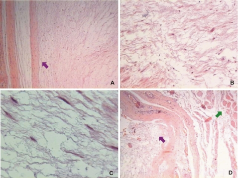
Myxoma is a neoplasm of mesenchymal origin composed of undifferentiated stellate cells in a myxoid stroma. This tumor can develop in a variety of locations. Myxomas that arise from skeletal muscles are called intramuscular myxomas. They usually occur in large skeletal muscles. Only ten cases of these benign tumors involving the neck muscles were reported in literature. Of them, only three were located at the paraspinal muscles. A 64-year-old woman presented with occipital and neck pain over 5 years noted an expansive painful lesion located at posterior cervical region with progressive volume increase in the last 12 months. Image exams revealed a large mass located in the left posterior region of the neck in contact with the C2, C3 and C4 laminae with no invasion of the vertebrae. Tumor total removal was performed through normal muscle margins and the vertebral periosteum was scraped. The tumor was encapsulated, lobulated with a gray-white appearance. The histological examination yielded the diagnosis of intramuscular myxoma. Follow-up at 1 year showed complete resolution of preoperative symptoms and no evidence of local recurrence. In conclusion, although rare, intramuscular myxoma should be included in differential diagnosis of cervical paraspinal tumors. We reported the fourth case of intramuscular myxoma in the paraspinal musculature of the neck. Despite its benign characteristics, local recurrence was reported after subtotal resection. Tumor total removal should be the goal of surgery.


Figures

Fig. 1
Fig. 1
Clinical appearance of the posterior cervical mass reveals a 6 × 6-cm fixed mass in the left posterior aspect of the neck
Fig. 2
Fig. 2
Cervical CT scan shows a septated, well-defined and hypodense mass in the left posterior neck. The mass does not enhance with intravenous contrast medium (a). Sagittal and axial T1-weighted MRI shows an intermediate to low signal (b) and the T2-weighted shows an increased signal (c) in the septaded cystic-looking mass
Fig. 3
Fig. 3
Intraoperative aspect of the tumor, which was encapsuleted with good cleavage plan (a). Tumor total removal was performed through normal muscle margins and the vertebral periosteum was scraped (b). Macroscopically cut surface of specimen is ovoid, graywish and gelatinous (c)
Fig. 4
Fig. 4
Histopathologic views of the intramuscular myxoma. a Photomicrography shows the capsulae of the tumor (purple set) (hematoxylin–eosin; ×4). b Low magnification appearance of the tumor characterized by a paucity of cells, abundance of mucoid material, and almost complete absence of vascular structures (hematoxylin–eosin; ×10). c Photomicrography at higher magnification reveals a myxoid stroma sparsely populated by small spindle shape cells and stellate cells without nuclear atypia and fibers (hematoxylin–eosin; ×40). d Low magnification show the tumor (purple set) surrounded by normal skeletal muscle (green set) (hematoxylin–eosin; ×4)

Similar articles

Cited by

References

    1. Abdelwahab AF, Kenan S, Hermann G, Lewis MM, Klein MJ. Intramuscular myxoma: magnetic resonance features. Br J Radiol. 1992;65:485–490. doi: 10.1259/0007-1285-65-774-485. - DOI - PubMed
    1. Allen PW. Myxoma is not a single entity: a review of the concept of myxoma. Ann Diagn Pathol. 2000;4:99–123. doi: 10.1016/S1092-9134(00)90019-4. - DOI - PubMed
    1. Andrews T, Kountakis SE, Maillard AA. Myxomas of the head and neck. Am J Otolaryngol. 2000;21:184–189. doi: 10.1016/S0196-0709(00)85022-X. - DOI - PubMed
    1. Canalis RF, Smith GA, Konrad HR. Myxomas of the head and neck. Arch Otolaryngol. 1976;102:300–305. - PubMed
    1. Crankson SJ, Al Namshan M, Al Mane K, Bamefleh H. Intramuscular myxoma: a rare neck mass in a child. Pediatr Radiol. 2002;32:120–122. doi: 10.1007/s00247-001-0603-5. - DOI - PubMed