. 2009 Apr;238(4):875-86.
doi: 10.1002/dvdy.21910.

Kit and foxd3 genetically interact to regulate melanophore survival in zebrafish

Affiliations

Kit and foxd3 genetically interact to regulate melanophore survival in zebrafish

Cynthia D Cooper et al. Dev Dyn. 2009 Apr.

Abstract

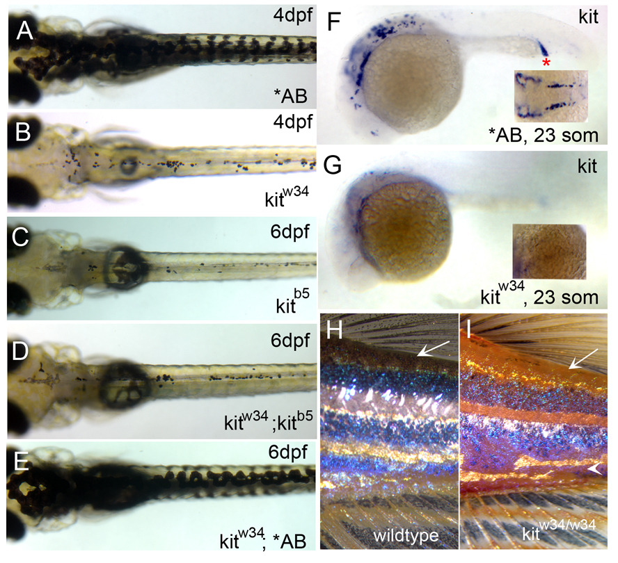
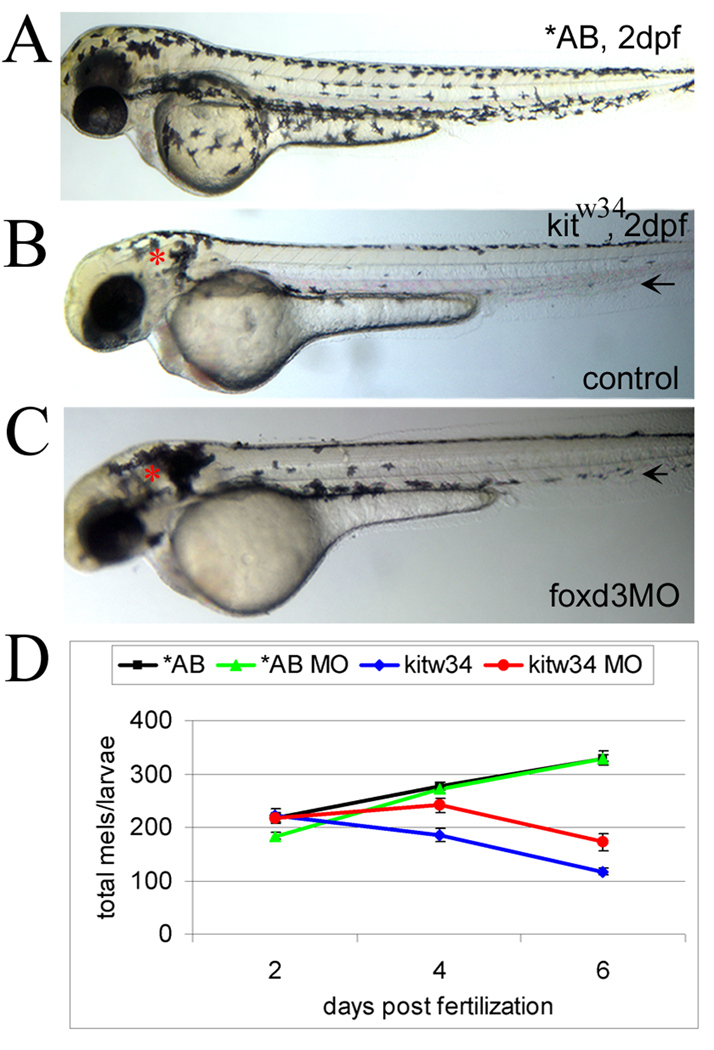
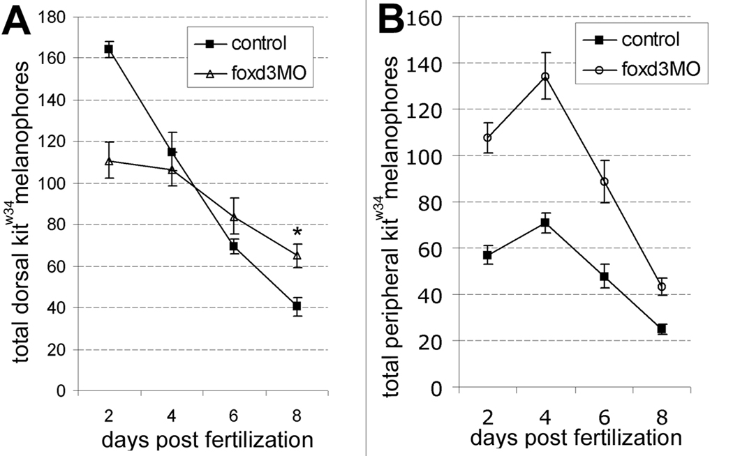
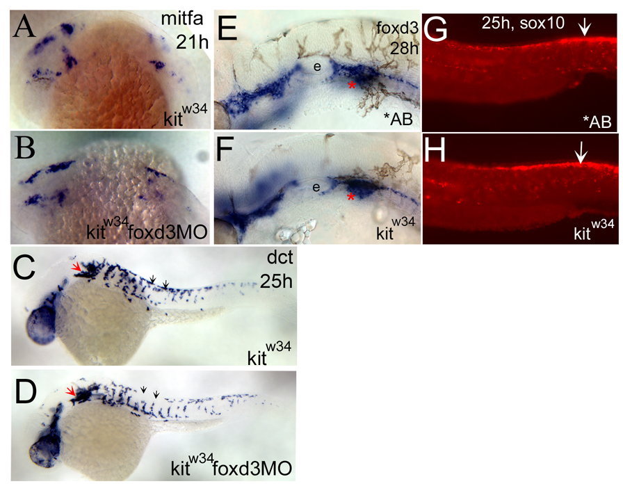
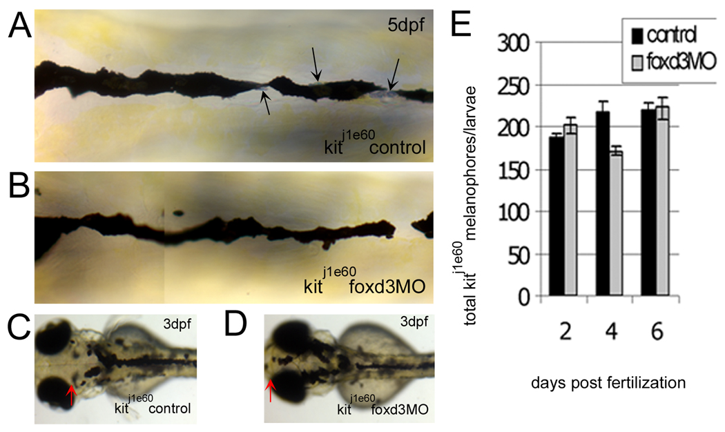
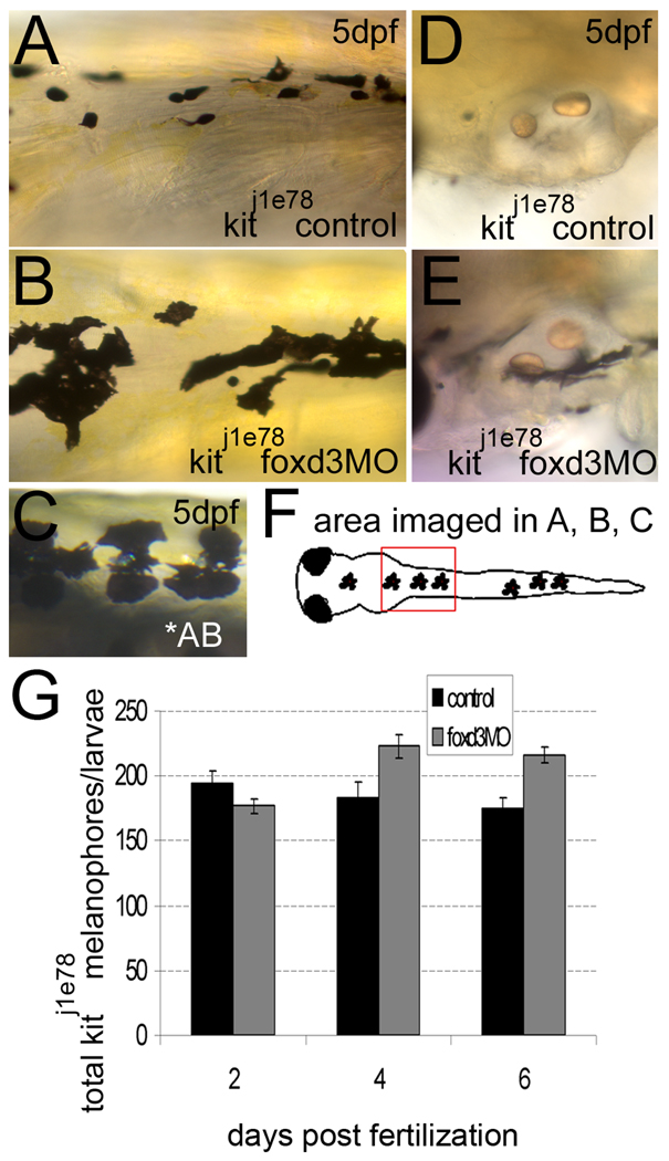
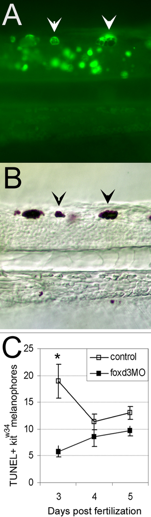
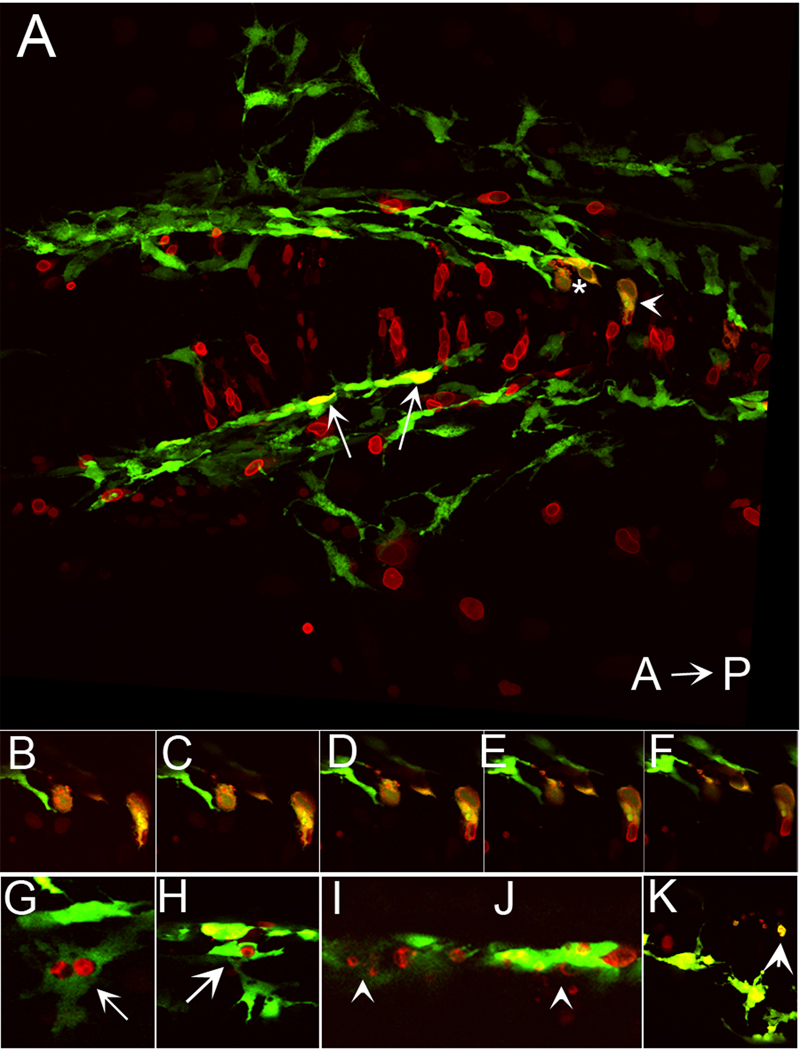
We have investigated the role of foxd3 activity in conjunction with signaling by the kit tyrosine kinase receptor in zebrafish black pigment cell (melanophore) development. As loss-of-function of these molecules individually has distinct effects on melanophore number, we have examined the phenotype of double mutants. Individuals with a null mutation in kit have fewer melanophores than wild-type, with cells lost through death. When kit mutants are injected with foxd3 antisense morpholino oligonucleotides or crossed with a foxd3 zebrafish mutant, they have more melanophores than their uninjected or foxd3+ counterparts. Examination of foxd3 loss-of-function in two additional kit mutants that differentially alter kit-dependent migration and survival indicates a change in melanophore number in survival mutants only. Consistently, TUNEL (terminal deoxynucleotidyl transferase-mediated deoxyuridinetriphosphate nick end-labeling) analysis confirms a partial rescue of melanophores from cell death. Ectopic expression of foxd3 indicates that foxd3 promotes early melanophore death only when kit is inactive. Taken together, these data suggest a kit-dependent role for foxd3 in the regulation of melanophore survival.


Figures

Fig. 1
Fig. 1
Characterization of kitw34 mutant larvae. (A) AB zebrafish at 4dpf, illustrating wildtype melanophore morphology and patterning. (B) kitw34 zebrafish at 4dpf, have small, rounded melanophores. Lateral and ventral stripe melanophores are largely missing by this stage. (C–E) kitw34/kitb5 complementation analysis. (C) In kitb5, 6dpf zebrafish, the majority of melanophores have died. (D) While progeny from a homozygous kitb5 and kitw34 cross show defects in melanophore morphology and survival, progeny from a kitw34 and AB cross are rescued and show wildtype melanophore patterning (E). (F, G) In situ analysis for kit mRNA during early melanogenesis. (F) Wildtype zebrafish express kit in the head, anterior trunk and intermediate cell mass (red asterisk) at 23 somites. (G) kitw34 embryos lack kit expression. H, I) kitw34 adults have a reduction in melanophores. (H) Wildtype pigment pattern showing three distinct dark stripes on the flank near the dorsal and caudal fins. (I) kitw34 adult showing reduced overall pigmentation, especially in dorsal areas (arrow) and throughout the dark stripes. The darks stripes are disorganized and the ventral stripe is lost with kit loss-of-function (arrowhead).
Fig. 2
Fig. 2
foxd3 loss-of-function reduces loss of melanophores in kitw34 zebrafish (A) In 2 dpf wildtype zebrafish (AB), melanophores have migrated extensively, anteriorly to the head and ventrally over the yolk. (B) In 2 dpf kitw34 zebrafish, melanophores are specified, but largely fail to migrate from sites of origin: the dorsal trunk and area caudal to the otic vesicle (red asterisk). (C) kitw34 zebrafish injected with foxd3 MO have more melanophores, especially apparent near the otic vesicle (foxd3MO, red asterisk). Both the ventral and lateral stripes show an increase in melanophores (black arrows). (D) Line graph showing counts of total melanophores in 2, 4 and 6dpf AB or kitw34 larvae, either uninjected (control) or injected with foxd3 MO. A significant change in the number of melanophores is observed in kitw34 mutants after injection of foxd3 MO (red) as compared to kitw34 uninjected controls (blue; p<0.0001 by 2-way ANOVA; 7–11 fish per time point), whereas AB foxd3 morphants (green) show no significant difference as compared to uninjected AB controls (black; p=0.49 by 2-way ANOVA; 9–13 fish per time point).
Fig. 3
Fig. 3
foxd3 loss-of-function alters the number of melanophores localized to the dorsal stripe and periphery. A) In the dorsal stripe, kitw34 control embryos (solid squares) lose melanophores quickly, showing a dramatic reduction by 8 dpf. Conversely, melanophores persist in kitw34 foxd3 morphants at higher numbers (*, p<0.05 by Bonferroni posttest at 8 dpf). Note that dorsal melanophores are initially present at lower numbers in kitw34 animals injected with foxd3 MO than in controls, but are present at higher numbers at 2 dpf in the periphery. B) The few melanophores detected in kitw34 lateral and ventral stripes (solid squares) at 2 dpf initially increase, and then steadily decline with each subsequent time point. Peripheral melanophores are detected at significantly higher numbers in 2 dpf in kitw34 foxd3 morphants as compared to uninjected kitw34 controls at all time periods (p<0.05 a by Bonferroni posttest).
Fig. 4
Fig. 4
foxd3 loss-of-function does not affect the number of melanoblasts specified from kitw34 neural crest. A,B) mitfa expression is detected in the head and anterior trunk of kitw34 21hpf embryos. This pattern is not dramatically altered in kitw34 foxd3 morphants. C, D) In 25 hpf kitw34 embryos, dopachrome tautomerase (dct) positive cells are found caudal to the eye and ear (red arrow), and in streams migrating ventrally between somites from the dorsal trunk (black arrows). kitw34 foxd3 morphants show some differences in migration (compare C and D, black arrows) but the overall number of cells appears similar. E, F) 28hpf AB and kitw34 embryos, processed by in situ hybridization for foxd3 message. foxd3 expression appears unchanged by kit loss-of-function. e; developing ear or otic vesicle G, H) 25hpf AB and kitw34 embryos, processed by fluorescent in situ hybridization for sox10 message. sox10 expression in premigratory neural crest cells is mostly unchanged (white arrows), although there is some reduction in peripheral expression, consistent with the reduction in melanoblast migration observed in kitw34 mutants.
Fig. 5
Fig. 5
The increase in kit mutant melanophores with foxd3 loss-of-function is not due to transfating of other foxd3 dependent cell types. A-D) kitj1e60 melanophore migration mutants were injected (or left uninjected; control) with foxd3 MO (foxd3MO), imaged at 3 (C, D) and 5dpf (A, B) and fixed at 2, 4 and 6 dpf for melanophore quantification. A) Melanophores localized to the anterior dorsal trunk display relatively normal, non-apoptotic morphology. Black arrows indicate silver pigment cells, iridophores. B) In kitj1e60 foxd3 morphants, foxd3 dependent iridophores are gone indicating MO function (average control and morphant iridophores ± standard deviation at 4dpf are 52±3 and 18±14, respectively), yet melanophore morphology and number remain similar to uninjected controls. C) kitj1e60 control melanophores do not localize to the anterior most portion of the head, indicating a migration defect. D) The presence of melanophores over the head is partially rescued with foxd3 loss-of-function (red arrows indicate anterior extent of melanophores). E) kitj1e60 melanophores are not significantly increased with foxd3 loss-of-function at 2, 4 and 6 dpf (p=0.31 by 2-way ANOVA; 8–15 fish per time point).
Fig. 6
Fig. 6
foxd3 loss-of-function improves the number and morphology of kit j1e78 survival mutant melanophores. A, B, D, E) kitj1e78 survival mutants were injected with foxd3 MO (foxd3MO) or uninjected embryos were examined as controls. A) Control kitj1e78 melanophores are small and rounded, characteristic of apoptosing melanophores. B) loss of foxd3 activity restores some wildtype morphology to kitj1e78 melanophores, and increases the number of melanophores (compare 6B to AB wildtype melanophores in 6C). D) kitj1e78 controls lack ventral stripe melanophores near the otic vesicle, which have apoptosed and been removed from the fish. E) kitj1e78 foxd3 morphants show increases in melanophores at this location, suggesting increased survival. F) Cartoon illustrating the location of images taken in A–C. G) Quantification at 2, 4 and 6 dpf of kitj1e78 and kitj1e78 foxd3 morphant melanophores. There is a statistically significant increase in kitj1e78 animals after foxd3 MO injection (p<0.005 by 2-way ANOVA; 10–19 fish per time point).
Fig. 7
Fig. 7
foxd3 loss-of-function partially rescues melanophores from apoptosis in kitw34 larvae. A) 3dpf kitw34 larvae analyzed by TUNEL assay. Examples of posterior trunk melanophores positive for TUNEL signal are marked with white arrowheads. B) Brightfield image of larvae shown in A. TUNEL positive melanophores (black arrowheads) are distinct in size and shape. C) Line graph indicating the total number of TUNEL positive melanophores found in the dorsal stripe of uninjected or foxd3 MO injected kitw34 larvae quantified at 3, 4 and 5 dpf. There is a significant decrease in TUNEL positive melanophores at 3 dpf with foxd3 loss-of-function. (control n=45 fish, 649 TUNEL+ cells; foxd3MO n=64 fish, 493 TUNEL+ cells; *, p<0.001 by Bonferroni posttest at 3dpf).
Fig. 8
Fig. 8
Ectopic expression of foxd3 promotes cell death in kit mutants. A) Confocal stack image showing a dorsal view of posterior head of 28 hpf kitw34/w34 embryo transgenic for mi::gfp and also expressing myc-foxd3 (red) under the control of a heat shock promoter. Expression of myc-foxd3 was induced at 19 hpf. Both wildtype (arrows) and fragmenting (asterisk and arrowhead) melanophores are observed. B- F) Confocal slices showing fragmenting cells indicated by asterisk and arrowhead in A. Note the presence of Foxd3 positive fragments within the GFP+ melanophores. G–H) Confocal slices showing additional examples of GFP/Foxd3+ cells designated as normal melanophores. I–J) Confocal slices showing examples of GFP/Foxd3+ cells designated as fragmenting melanophores (arrowheads).

References

    1. Alkhateeb A, Fain PR, Spritz RA. Candidate functional promoter variant in the FOXD3 melanoblast developmental regulator gene in autosomal dominant vitiligo. Journal of Investigative Dermatology. 2005;125:388–391. - PubMed
    1. Bernex F, De Sepulveda P, Kress C, Elbaz C, Delouis C, Panthier JJ. Spatial and temporal patterns of c-kit-expressing cells in WlacZ/+ and WlacZ/WlacZ mouse embryos. Development. 1996;122:3023–3033. - PubMed
    1. Blechman JM, Lev S, Brizzi MF, Leitner O, Pegoraro L, Givol D, Yarden Y. Soluble c-kit proteins and antireceptor monoclonal antibodies confine the binding site of the stem cell factor. Journal of Biological Chemistry. 1993;268:4399–4406. - PubMed
    1. Cooper CD, Raible DW. Mechanisms for reaching the differentiated state: Insights from neural crest derived melanocytes. Sem Cell Dev Biol 2008. 2008 Sep 30; [Epub ahead of print] - PMC - PubMed
    1. Copeland NG, Gilbert DJ, Cho BC, Donovan PJ, Jenkins NA, Cosman D, Anderson D, Lyman SD, Williams DE. Mast cell growth factor maps near the steel locus on mouse chromosome 10 and is deleted in a number of steel alleles. Cell. 1990;63:175–183. - PubMed

Publication types

MeSH terms

LinkOut - more resources