

Neuroprotective role of erythropoietin by antiapoptosis in the retina
- PMID: 19301424
- PMCID: PMC5161804
- DOI: 10.1002/jnr.22046
Neuroprotective role of erythropoietin by antiapoptosis in the retina
Abstract
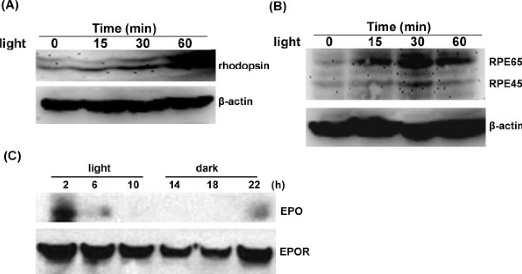
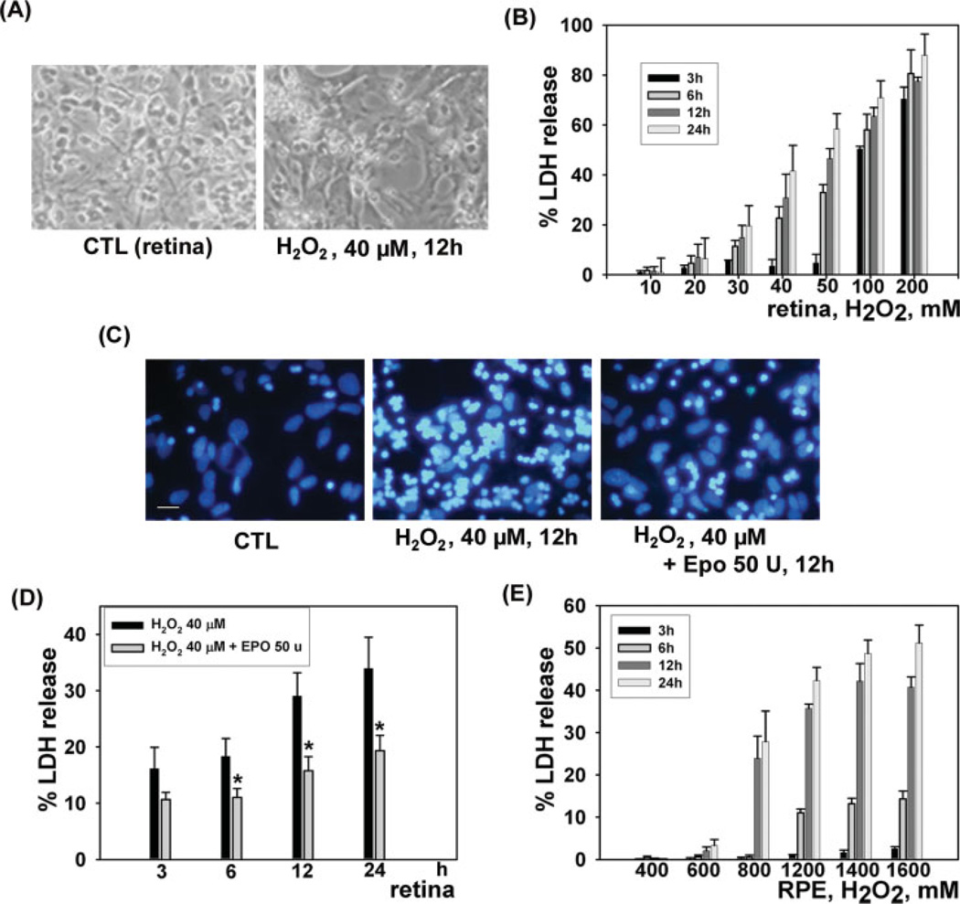
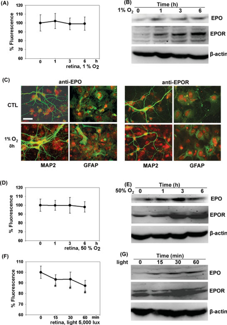
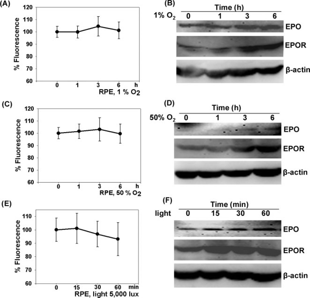
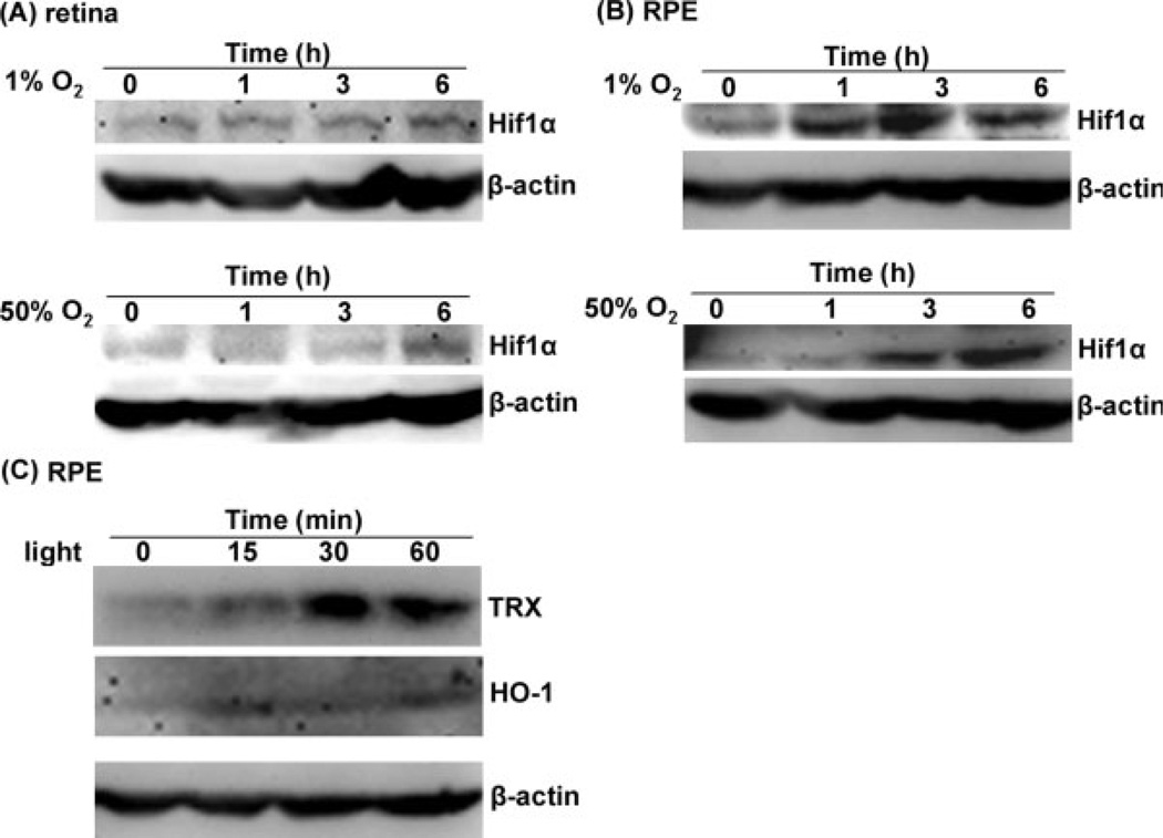
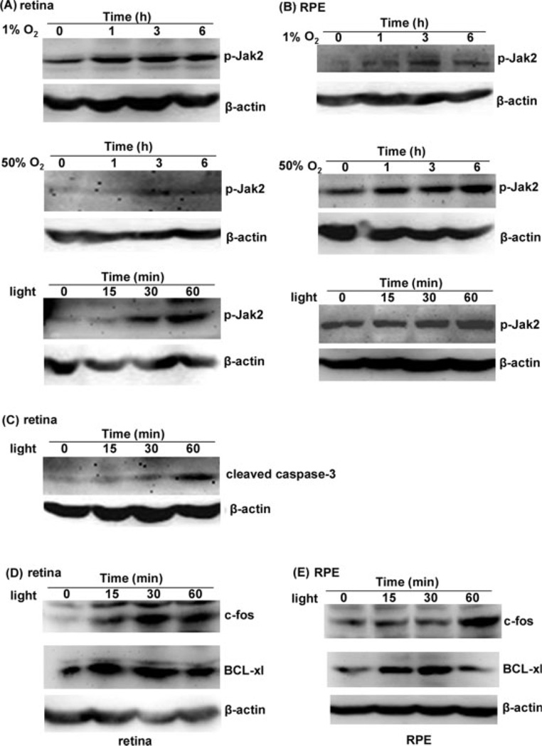
Erythropoietin (EPO) stimulates red blood cell production, in part by inhibiting apoptosis of the red blood cell precursors. The erythropoietic effects of EPO are circadian stage dependent. Retinal injury due to light occurs through oxidative mechanisms and is manifest by retinal and retinal pigment epithelium (RPE) cells apoptosis. The visual cycle might be circadian coordinated as a means of effectively protecting the retina from the detrimental effects of light-induced, oxygen-dependent, free radical-mediated damage, especially at the times of day when light is more intense. We show that the retinal expression of EPO and its receptor (EPOR), as well as subsequent Janus kinase 2 (Jak2) phosphorylations, are each tightly linked to a specific time after oxidative stress and in anticipation of daily light onset. This is consistent with physiological protection against daily light-induced, oxidatively mediated retinal apoptosis. In vitro, we verify that EPO protects RPE cells from light, hyperoxia, and hydrogen peroxide-induced retinal cell apoptosis, and that these stimuli increase EPO and EPOR expression in cultured RPE cells. Together, these data support the premise that EPO and its EPOR interactions represent an important retinal shield from physiologic and pathologic light-induced oxidative injury.
Copyright 2009 Wiley-Liss, Inc.
Figures
Similar articles
-
Erythropoietin is a paracrine mediator of ischemic tolerance in the brain: evidence from an in vitro model.J Neurosci. 2002 Dec 1;22(23):10291-301. doi: 10.1523/JNEUROSCI.22-23-10291.2002. J Neurosci. 2002. PMID: 12451129 Free PMC article.
-
Neuroprotective effects of erythropoietin against sevoflurane-induced neuronal apoptosis in primary rat cortical neurons involving the EPOR-Erk1/2-Nrf2/Bach1 signal pathway.Biomed Pharmacother. 2017 Mar;87:332-341. doi: 10.1016/j.biopha.2016.12.115. Epub 2017 Jan 5. Biomed Pharmacother. 2017. PMID: 28064106
-
Erythropoietin ameliorates early brain injury after subarachnoid haemorrhage by modulating microglia polarization via the EPOR/JAK2-STAT3 pathway.Exp Cell Res. 2017 Dec 15;361(2):342-352. doi: 10.1016/j.yexcr.2017.11.002. Epub 2017 Nov 2. Exp Cell Res. 2017. PMID: 29102603
-
Erythropoietin and the hypoxic brain.J Exp Biol. 2004 Aug;207(Pt 18):3233-42. doi: 10.1242/jeb.01049. J Exp Biol. 2004. PMID: 15299044 Review.
-
Erythropoietin action in stress response, tissue maintenance and metabolism.Int J Mol Sci. 2014 Jun 10;15(6):10296-333. doi: 10.3390/ijms150610296. Int J Mol Sci. 2014. PMID: 24918289 Free PMC article. Review.
Cited by
-
Erythropoietin levels in aqueous humor of patients with glaucoma.Mol Vis. 2012;18:1991-5. Epub 2012 Jul 18. Mol Vis. 2012. PMID: 22876126 Free PMC article.
-
Dual Switch Mechanism of Erythropoietin as an Antiapoptotic and Pro-Angiogenic Determinant in the Retina.ACS Omega. 2020 Aug 12;5(33):21113-21126. doi: 10.1021/acsomega.0c02763. eCollection 2020 Aug 25. ACS Omega. 2020. PMID: 32875248 Free PMC article.
-
Revisiting the role of erythropoietin for treatment of ocular disorders.Eye (Lond). 2016 Oct;30(10):1293-1309. doi: 10.1038/eye.2016.94. Epub 2016 Jun 10. Eye (Lond). 2016. PMID: 27285322 Free PMC article. Review.
-
Human erythropoietin effect in postoperative visual loss following spine surgery: a case report.Anesth Pain Med. 2014 Apr 6;4(2):e7291. doi: 10.5812/aapm.7291. eCollection 2014 May. Anesth Pain Med. 2014. PMID: 24790903 Free PMC article.
-
Disruption of Angiogenesis by Anthocyanin-Rich Extracts of Hibiscus sabdariffa.Int J Sci Eng Res. 2017 Feb;8(2):299-307. doi: 10.14299/ijser.2017.02.009. Int J Sci Eng Res. 2017. PMID: 28459020 Free PMC article.
References
-
- Bellamy WT, Alberts DS, Dorr RT. Daily variation in non-protein sulfhydryl levels of human bone marrow. Eur J Cancer Clin Oncol. 1988;24:1759–1762. - PubMed
-
- Böcker-Meffert S, Rosenstiel P, Röhl C, Warneke N, Held-Feindt J, Sievers J, Lucius R. Erythropoietin and VEGF promote neural outgrowth from retinal explants in postnatal rats. Invest Ophthalmol Vis Sci. 2002;43:2021–2026. - PubMed
-
- Brines M, Cerami A. Emerging biological roles for erythropoietin in the nervous system. Nat Rev Neurosci. 2005;6:484–494. - PubMed
-
- Calapai G, Marciano MC, Corica F, Allegra A, Parisi A, Frisina N, Caputi AP, Buemi M. Erythropoietin protects against brain ischemic injury by inhibition of nitric oxide formation. Eur J Pharmacol. 2000;401:349–356. - PubMed
MeSH terms
Substances
Grants and funding
LinkOut - more resources
Full Text Sources
Medical
Research Materials
Miscellaneous
