Review
. 2009:27:339-62.
doi: 10.1146/annurev.immunol.021908.132554.

The ins and outs of leukocyte integrin signaling

Affiliations
Review

The ins and outs of leukocyte integrin signaling

Clare L Abram et al. Annu Rev Immunol. 2009.

Abstract

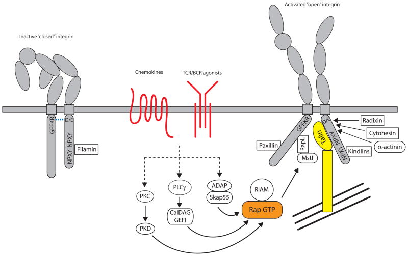
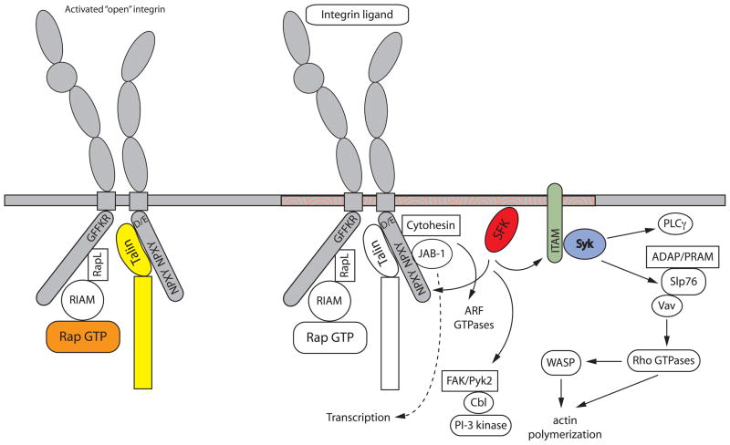
Integrins are the principal cell adhesion receptors that mediate leukocyte migration and activation in the immune system. These receptors signal bidirectionally through the plasma membrane in pathways referred to as inside-out and outside-in signaling. Each of these pathways is mediated by conformational changes in the integrin structure. Such changes allow high-affinity binding of the receptor with counter-adhesion molecules on the vascular endothelium or extracellular matrix and lead to association of the cytoplasmic tails of the integrins with intracellular signaling molecules. Leukocyte functional responses resulting from outside-in signaling include migration, proliferation, cytokine secretion, and degranulation. Here, we review the key signaling events that occur in the inside-out versus outside-in pathways, highlighting recent advances in our understanding of how integrins are activated by a variety of stimuli and how they mediate a diverse array of cellular responses.


Figures

Figure 1
Figure 1
Alignment of transmembrane and cytoplasmic regions of leukocyte integrins. Data is taken from UniProt (www.uniprot.org), and amino acid numbering does not include the signal sequence. The solid orange box indicates the transmembrane domain predicted by the Hidden Markov model (121). The open red box indicates residues that were experimentally determined to be within the lipid bilayer (122). Residues boxed in yellow are important for the salt bridge linking the α and β integrin tails. The purple boxes indicate the two NPXY/F motifs in the β integrin tails.
Figure 2
Figure 2
Integrin inside-out signaling. The figure outlines the key signaling events that occur downstream of chemokine, T and B cell receptors that lead to integrin activation. Inactive integrins exist in a bent conformation and the α and β cytoplasmic tails are held in close proximity by a salt bridge between residues found in the membrane proximal region of the tail. Activation of a variety of signaling pathways results in the recruitment of GTP-bound Rap1 and activated talin to the integrin, leading to tail separation. The conformational change in the cytoplasmic region is transmitted through the integrin transmembrane domain and results in structural changes in the extracellular region, leading to an open conformation that can bind ligand with high affinity. The C-terminal rod domain of talin interacts with the actin cytoskeleton, to provide physical coupling of the integrin to the actin network of the cell. Many other molecules have been shown to interact with integrin cytoplasmic tails, but exactly how these interactions are coordinated with integrin activation is unclear.
Figure 3
Figure 3
Integrin outside-in signaling. The figure details integrin-mediated signaling events that occur downstream of ligand binding. Zhu et al have shown that outside-in signaling requires structural changes with the cytoplasmic region of integrin tails (87). Activation of Src family kinases is a key step, although the exact mechanism by which this occurs is unclear, and results in phosphorylation of a variety of downstream molecules. These include ITAM-containing adapters that, when phosphorylated, lead to the recruitment and activation of Syk or ZAP-70 kinases. These kinases in turn phosphorylate various substrates including SLP76 and Vav. This lead us to propose that integrin outside-in signaling is analogous to signaling downstream of immunoreceptors, as indicated by the molecules in bold (85, 86). Vav activates Rho GTPases leading to actin cytoskeletal reorganization. Src family kinases can also activate FAK and Pyk2 kinases leading to Cbl phosphorylation and recruitment, as well as PI3K activation. Association of other molecules such as JAB and cytohesin with the integrin cytoplasmic tails activates other downstream signaling pathways. In this figure, signaling is depicted as happening in lipid rafts (indicated as orange swirls in the membrane), although the role of rafts in integrin signaling differs in various cell types.

Similar articles

Cited by

References

    1. Ley K, Laudanna C, Cybulsky MI, Nourshargh S. Getting to the site of inflammation: the leukocyte adhesion cascade updated. Nat Rev Immunol. 2007;7:678–89. - PubMed
    1. Luo BH, Carman CV, Springer TA. Structural basis of integrin regulation and signaling. Annu Rev Immunol. 2007;25:619–47. - PMC - PubMed
    1. Arnaout MA, Goodman SL, Xiong JP. Structure and mechanics of integrin-based cell adhesion. Curr Opin Cell Biol. 2007;19:495–507. - PMC - PubMed
    1. Kinashi T. Intracellular signalling controlling integrin activation in lymphocytes. Nat Rev Immunol. 2005;5:546–59. - PubMed
    1. McLeod SJ, Shum AJ, Lee RL, Takei F, Gold MR. The Rap GTPases regulate integrin-mediated adhesion, cell spreading, actin polymerization, and Pyk2 tyrosine phosphorylation in B lymphocytes. J Biol Chem. 2004;279:12009–19. - PubMed

Publication types

MeSH terms

LinkOut - more resources