

Magnetic resonance microscopy defines ethanol-induced brain abnormalities in prenatal mice: effects of acute insult on gestational day 8
- PMID: 19302087
- PMCID: PMC2748865
- DOI: 10.1111/j.1530-0277.2009.00921.x
Magnetic resonance microscopy defines ethanol-induced brain abnormalities in prenatal mice: effects of acute insult on gestational day 8
Abstract
Background: Magnetic resonance microscopy (MRM), magnetic resonance imaging (MRI) at microscopic levels, provides unprecedented opportunities to aid in defining the full spectrum of ethanol's insult to the developing brain. This is the first in a series of reports that, collectively, will provide an MRM-based atlas of developmental stage-dependent structural brain abnormalities in a Fetal Alcohol Spectrum Disorders (FASD) mouse model. The ethanol exposure time and developmental stage examined for this report is gestational day (GD) 8 in mice, when the embryos are at early neurulation stages; stages present in humans early in the fourth week postfertilization.
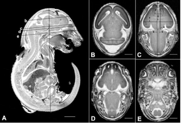
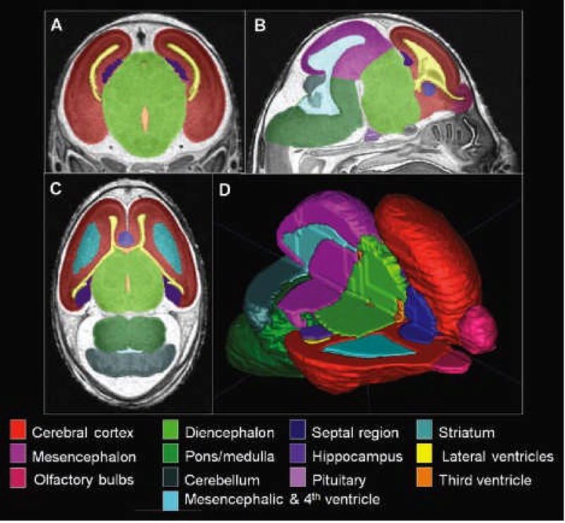
Methods: For this study, pregnant C57Bl/6J mice were administered an ethanol dosage of 2.8 g/kg intraperitoneally at 8 days, 0 hour and again at 8 days, 4 hours postfertilization. On GD 17, fetuses that were selected for MRM analyses were immersion fixed in a Bouin's/Prohance solution. Control fetuses from vehicle-treated dams were stage-matched to those that were ethanol-exposed. The fetal mice were scanned ex vivo at 7.0 T and 512 x 512 x 1024 image arrays were acquired using 3-D spin warp encoding. The resulting 29 microm (isotropic) resolution images were processed using ITK-SNAP, a 3-D segmentation/visualization tool. Linear and volume measurements were determined for selected brain, head, and body regions of each specimen. Comparisons were made between control and treated fetuses, with an emphasis on determining (dis)proportionate changes in specific brain regions.
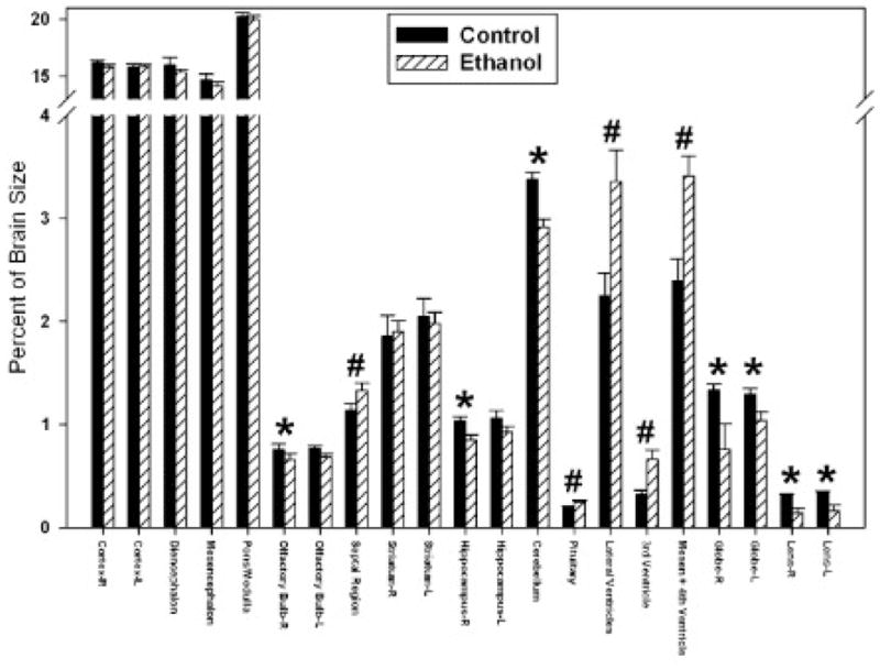
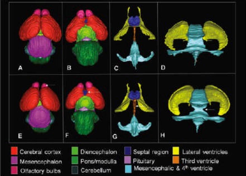
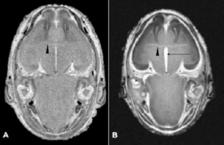
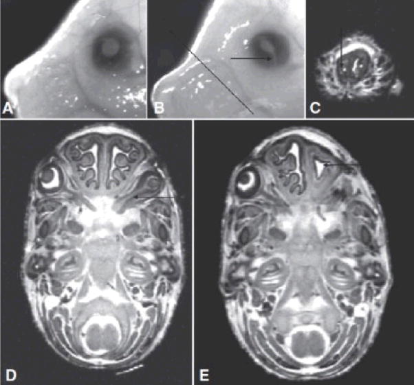
Results: As compared with controls, the crown-rump lengths of stage-matched ethanol-exposed GD 17 fetuses were significantly reduced, as were brain and whole body volumes. Volume reductions were notable in every brain region examined, with the exception of the pituitary and septal region, and were accompanied by increased ventricular volumes. Disproportionate regional brain volume reductions were most marked on the right side and were significant for the olfactory bulb, hippocampus, and cerebellum; the latter being the most severely affected. Additionally, the septal region and the pituitary were disproportionately large. Linear measures were consistent with those of volume. Other dysmorphologic features noted in the MR scans were choanal stenosis and optic nerve coloboma.
Conclusions: This study demonstrates that exposure to ethanol occurring in mice at stages corresponding to the human fourth week postfertilization results in structural brain abnormalities that are readily identifiable at fetal stages of development. In addition to illustrating the utility of MR microscopy for analysis of an FASD mouse model, this work provides new information that confirms and extends human clinical observations. It also provides a framework for comparison of structural brain abnormalities resulting from ethanol exposure at other developmental stages and dosages.
Figures
Similar articles
-
Magnetic resonance microscopy-based analyses of the brains of normal and ethanol-exposed fetal mice.Birth Defects Res A Clin Mol Teratol. 2010 Nov;88(11):953-64. doi: 10.1002/bdra.20719. Epub 2010 Sep 14. Birth Defects Res A Clin Mol Teratol. 2010. PMID: 20842647 Free PMC article.
-
Magnetic resonance microscopy defines ethanol-induced brain abnormalities in prenatal mice: effects of acute insult on gestational day 7.Alcohol Clin Exp Res. 2010 Jan;34(1):98-111. doi: 10.1111/j.1530-0277.2009.01071.x. Epub 2009 Oct 23. Alcohol Clin Exp Res. 2010. PMID: 19860813 Free PMC article.
-
Maternal oral intake mouse model for fetal alcohol spectrum disorders: ocular defects as a measure of effect.Alcohol Clin Exp Res. 2006 Oct;30(10):1791-8. doi: 10.1111/j.1530-0277.2006.00212.x. Alcohol Clin Exp Res. 2006. PMID: 17010146
-
Genesis of alcohol-induced craniofacial dysmorphism.Exp Biol Med (Maywood). 2005 Jun;230(6):366-75. doi: 10.1177/15353702-0323006-04. Exp Biol Med (Maywood). 2005. PMID: 15956766 Review.
-
Teratogenic actions of ethanol in the mouse: a minireview.Pharmacol Biochem Behav. 1996 Dec;55(4):501-13. doi: 10.1016/s0091-3057(96)00255-9. Pharmacol Biochem Behav. 1996. PMID: 8981580 Review.
Cited by
-
Low and moderate prenatal ethanol exposures of mice during gastrulation or neurulation delays neurobehavioral development.Neurotoxicol Teratol. 2015 Sep-Oct;51:1-11. doi: 10.1016/j.ntt.2015.07.003. Epub 2015 Jul 11. Neurotoxicol Teratol. 2015. PMID: 26171567 Free PMC article.
-
Pituitary lacks sexual dimorphism and displays reduced signal intensity on T1-weighted MRI in adolescents with histories of heavy prenatal alcohol exposure.Neurotoxicol Teratol. 2016 Sep-Oct;57:106-111. doi: 10.1016/j.ntt.2016.09.001. Epub 2016 Sep 9. Neurotoxicol Teratol. 2016. PMID: 27616668 Free PMC article.
-
Detecting Neurodevelopmental Effects of Early-Gestation Ethanol Exposure: A Nonhuman Primate Model of Ethanol Drinking During Pregnancy.Alcohol Clin Exp Res. 2019 Feb;43(2):250-261. doi: 10.1111/acer.13938. Epub 2019 Jan 11. Alcohol Clin Exp Res. 2019. PMID: 30549282 Free PMC article.
-
High-resolution imaging in studies of alcohol effect on prenatal development.Adv Drug Alcohol Res. 2023;3:10790. doi: 10.3389/adar.2023.10790. Epub 2023 Feb 1. Adv Drug Alcohol Res. 2023. PMID: 37593366 Free PMC article.
-
Distinct neurobehavioral dysfunction based on the timing of developmental binge-like alcohol exposure.Neuroscience. 2014 Nov 7;280:204-19. doi: 10.1016/j.neuroscience.2014.09.008. Epub 2014 Sep 18. Neuroscience. 2014. PMID: 25241068 Free PMC article.
References
-
- Abel EL. Fetal alcohol syndrome: a cautionary note. Curr Pharm Des. 2006;12:1521–1529. - PubMed
-
- Ammann AJ, Wara DW, Cowan MJ, Barrett DJ, Stiehm ER. The DiGeorge syndrome and the fetal alcohol syndrome. Am J Dis Child. 1982;136:906–908. - PubMed
-
- Autti-Rämö I, Autti T, Korkman M, Kettunen S, Salonen O, Valanne L. MRI findings in children with school problems who had been exposed prenatally to alcohol. Dev Med Child Neurol. 2002;44:98–106. - PubMed
-
- Bonthius DJ, West JR. Permanent neuronal deficits in rats exposed to alcohol during the brain growth spurt. Teratology. 1991;44:147–163. - PubMed
-
- Bookstein FL, Sampson PD, Connor PD, Streissguth AP. Midline corpus callosum is a neuroanatomical focus of fetal alcohol damage. Anat Rec. 2002;269:162–174. - PubMed
Publication types
MeSH terms
Substances
Grants and funding
LinkOut - more resources
Full Text Sources
Medical
Research Materials
