Review
. 2009 Mar;126(3):316-28.
doi: 10.1111/j.1365-2567.2008.03050.x.

Restricting activation-induced cytidine deaminase tumorigenic activity in B lymphocytes

Affiliations
Review

Restricting activation-induced cytidine deaminase tumorigenic activity in B lymphocytes

Rafael Casellas et al. Immunology. 2009 Mar.

Abstract

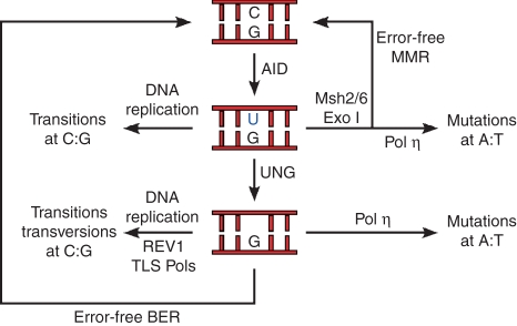
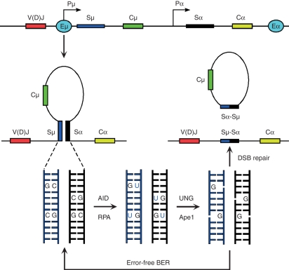
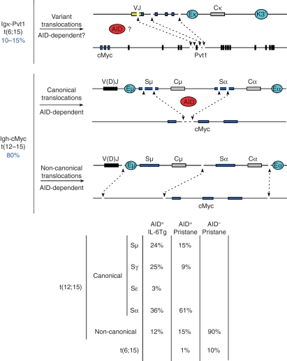
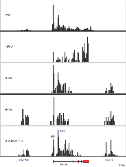
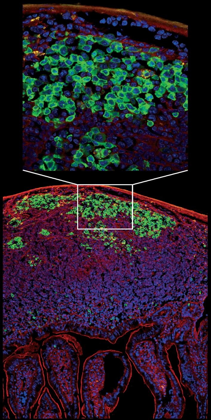
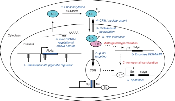
DNA breaks play an essential role in germinal centre B cells as intermediates to immunoglobulin class switching, a recombination process initiated by activation-induced cytidine deaminase (AID). Immunoglobulin gene hypermutation is likewise catalysed by AID but is believed to occur via single-strand DNA breaks. When improperly repaired, AID-mediated lesions can promote chromosomal translocations (CTs) that juxtapose the immunoglobulin loci to heterologous genomic sites, including oncogenes. Two of the most studied translocations are the t(8;14) and T(12;15), which deregulate cMyc in human Burkitt's lymphomas and mouse plasmacytomas, respectively. While a complete understanding of the aetiology of such translocations is lacking, recent studies using diverse mouse models have shed light on two important issues: (1) the extent to which non-specific or AID-mediated DNA lesions promote CTs, and (2) the safeguard mechanisms that B cells employ to prevent AID tumorigenic activity. Here we review these advances and discuss the usage of pristane-induced mouse plasmacytomas as a tool to investigate the origin of Igh-cMyc translocations and B-cell tumorigenesis.


Figures

Figure 1
Figure 1
Current model of somatic hypermutation (based on ref. 123). Activation-induced cytidine deiminase (AID)-mediated deamination of cytidines at immunoglobulin genes results in U : G mispairs. DNA replication over uracils will promote transition mutations (C/G to T/A). Uracil can also be repaired in an error-free fashion by either base excision repair (BER) or mismatch repair (MMR). If uracils are recognized by uracil-N-glycosylase 1 (UNG) in the context of DNA replication however, abasic sites would be repaired by REV1 and other translesion polymerases leading to both transition and transversions at C : Gs. Following replication, U : G mismatches or abasic sites would engage the activity of polymerase η (pol η) resulting in mutations primarily at A : T pairs.
Figure 2
Figure 2
Schematics representing class switch recombination from Sμ to Sα. Recombining switch domains are rendered accessible to activation-induced cytidine deiminase (AID) and replication protein A (RPA) by the concerted action of I-promoters and the μ (5′) and α (3′) enhancers (E). Removal of uracils by uracil-N-glycosylase 1 (UNG) and nicking of the DNA backbone by Ape nucleases results in the formation of staggered DNA breaks, which are either repaired in an error-free manner by base excision repair (BER) or serve as substrates for double-strand break (DSB) repair and/or recombination. Although not depicted, mismatch repair activity induces class switch recombination via a similar mechanism.
Figure 3
Figure 3
Classification of chromosomal translocations isolated from mouse plasmacytomas. Upper schematic exemplifies variant translocations [t(6;15)] that juxtapose VJk domains to Pvt1 exon 5. Middle schematic shows activation-induced cytidine deiminase (AID)-mediated deamination during class switch recombination results in the formation of DNA double-strand breaks at S domains and less frequently at cMyc intron 1. Aberrant repair of these lesions can lead to the juxtaposition of cMyc to the immunoglobulin loci. Lower schematic: although at a significantly lesser extent, random, AID-independent DNA breaks can also translocate the cMyc gene to the immunoglobulin loci leading to T(12;15). Translocation breakpoints in these non-canonical translocations often map several kilobases away from S domains or the cMyc gene itself. Right table indicates the percentage of CTs involving diverse S domains in AID+/+ interleukin-6 transgenic mice, and pristane-treated AID+/+ and AID−/− B/c mice.
Figure 4
Figure 4
Partial epigenetic map of the mouse Aicda locus (35, 400 bps) in lipopolysaccharide + interleukin-4 activated B cells depicting polymerase II binding, messenger RNA (mRNA) production, histone H3 and H4 acetylation, and histone H3 lysine 4 mono-, di-, or tri-methylation (H3K4me1-2-3). Phylogenetically conserved elements CNSVIII, activation-induced cytidine deiminase (AID) basal promoter (BP), AID intronic enhancer (AIDE), and CNSX are also highlighted. Mapping (200 base pair resolution) was produced by Solexa DNA microsequencing of immunoprecipitated chromatin (PolII, H3Ac, H4Ac, H3K4me1-2-3) or polyA+ isolated mRNA (Yamane and Casellas, manuscript in preparation).
Figure 5
Figure 5
Micrograph showing a longitudinal section of intestinal villi and Peyer’s patches from the jejunum of activation-induced cytidine deiminase-green fluorescent protein (AID-GFP) mice. A close-up view of the germinal centre reveals AID localization is mostly cytoplasmic. Cells were stained with DAPI (blue) and F-actin was labelled with phalloidin (red). The image was prepared by Susan Lim and collected by Kristien Zaal using a Leica SP5 confocal microscope.
Figure 6
Figure 6
Summary of activation-induced cytidine deiminase (AID) regulatory mechanisms: 1- Spatiotemporal targeting of AID gene transcription to activated and germinal centre B cells. This is achieved by the concerted action of transcription factors and the Aicda epigenetic landscape. 2- microRNA regulation of AID messenger RNA half-life. 3- Protein kinase A (PKA) and/or protein kinase C (PKC) -mediated AID phosphorylation. 4- AID nuclear export mediated by CRM1. 5- Nuclear proteasomal degradation. 5- AID interaction with single-stranded (ss) DNA binding protein replication protein A (RPA), which allows AID to deaminate transcribed ssDNA. 7- Preferential recruitment of the AID–RPA complex to immunoglobulin genes. 8- Mistargeted hypermutation to non-immunoglobulin genes like cMyc counteracted by error-free base excision repair (BER) and mismatch repair. 9- Immunoglobulin-oncogene translocations eliminated by apoptosis via P53 activation. This safeguard mechanism might also get rid of B lymphocytes carrying extensive DNA damage.

References

    1. Neuberger MS. Antibody diversification by somatic mutation: from Burnet onwards. Immunol Cell Biol. 2008;86:124–32. - PubMed
    1. Honjo T, Kinoshita K, Muramatsu M. Molecular mechanism of class switch recombination: linkage with somatic hypermutation. Annu Rev Immunol. 2002;20:165–96. - PubMed
    1. Revy P, Muto T, Levy Y, et al. Activation-induced cytidine deaminase (AID) deficiency causes the autosomal recessive form of the Hyper-IgM syndrome (HIGM2). [see comments] Cell. 2000;102:565–75. - PubMed
    1. Muramatsu M, Kinoshita K, Fagarasan S, Yamada S, Shinkai Y, Honjo T. Class switch recombination and hypermutation require activation-induced cytidine deaminase (AID), a potential RNA editing enzyme. [see comments] Cell. 2000;102:553–63. - PubMed
    1. Harris RS, Sale JE, Petersen-Mahrt SK, Neuberger MS. AID is essential for immunoglobulin V gene conversion in a cultured B cell line. Curr Biol. 2002;12:435–8. - PubMed

Publication types

MeSH terms

Substances