Review
. 2009 May;100(5):792-7.
doi: 10.1111/j.1349-7006.2009.01123.x. Epub 2009 Mar 15.

Cellular senescence: its role in tumor suppression and aging

Affiliations
Review

Cellular senescence: its role in tumor suppression and aging

Naoko Ohtani et al. Cancer Sci. 2009 May.

Abstract

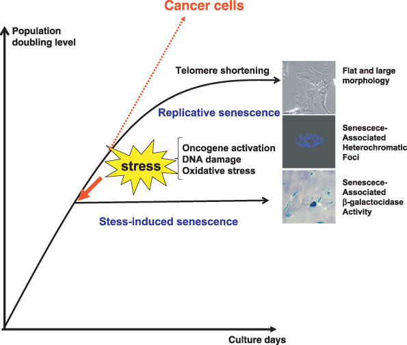
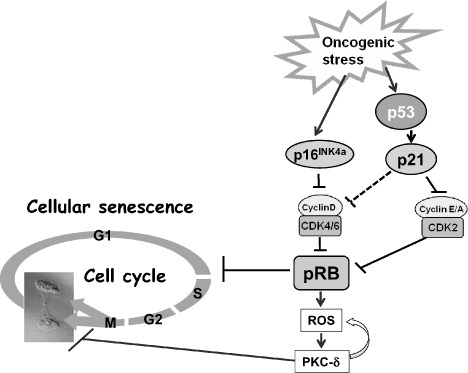
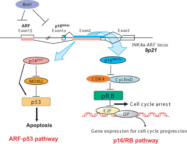
In normal tissue, cell division is carefully regulated to maintain the correct proliferative balance. Abnormal cell division underlies many hypoproliferative and hyperproliferative disorders, including cancer, and a better understanding of the mechanisms involved could lead to new strategies for treatment and prevention. Cellular senescence, a state of irreversible growth arrest, was first described as a limit to the replicative life span of somatic cells after serial cultivation in vitro. Recently, however, it has also been shown to be triggered prematurely by potentially oncogenic stimuli such as oncogene expression, oxidative stress, and DNA damage in cell culture studies. These data suggest that cellular senescence is therefore acting as a tumor-protective fail-safe mechanism. However, the significance of cellular senescence has remained an issue of debate over the years, with the possibility that it might be a cell culture-related artifact. Recent reports on oncogene-induced senescence detected in premalignant tumors have provided evidence to validate its role as a physiological response to prevent oncogenesis in vivo. In this review, we discuss the mechanisms for cellular senescence and its roles in vivo.


Figures

Figure 1
Figure 1
Cellular senescence. Normal human fibroblasts enter a state of irreversible growth arrest after a finite number of cell divisions in vitro caused by telomere shortening but cancer cells appear to bypass this replicative limit and proliferate indefinitely. Recent reports have shown that cellular senescence can also be induced prematurely by a number of cellular stresses such as oncogenic stimuli, oxidative stress, and DNA damage, before reaching their limits of replicative life span. This type of cellular senescence is called ‘stress‐induced senescence’. Senescent cells are characterized by a large and flat morphology, senescence‐associated acidic β‐galactosidase activity, and senescence‐associated heterochromatic foci.
Figure 2
Figure 2
Molecular mechanisms of cellular senescence. Oncogenic stress induces p16 and the p53‐target p21. When protein retinoblastoma (pRb) is fully activated by high‐level expression of p16INK4a, mitogenic signals, in turn, increase the level of reactive oxygen species (ROS) and elicit a positive feedback activation of the ROS–PKC‐δ signaling pathway. Elevated levels of p16INK4a therefore establish the autonomous activation of ROS–PKC‐δ signaling, leading to an irrevocable block to cytokinesis in human senescent cells. CDK, cyclin‐dependent kinase.
Figure 3
Figure 3
Ink4a/Arf locus. The p16INK4a gene is located in the Ink4a/Arf locus in human chromosome 9p21, and this locus encodes not only p16INK4a but also Arf via a shared second exon using a different translational reading frame. These two protein products participate in major tumor‐suppressor networks that are inactivated in human cancer. p16INK4a binds directly to and inhibits the activity of cyclin‐dependent kinase (CDK) 4 and CDK6 and hence activates the retinoblastoma (RB) tumor‐suppressor protein whereas ARF binds directly to mouse double minutes (MDM2) resulting in the stabilization and activation of the p53 tumor suppressor. This locus is repressed by polycomb proteins such as Bmi1 (B lymphoma Mo‐MLV insertion region 1). DP, DRTP1‐polypeptide‐1.
Figure 4
Figure 4
Real‐time in vivo imaging of p21Waf1/Cip1 gene expression after doxorubicin (DXR) treatment. We established a transgenic mouse line (p21‐p‐luc) expressing firefly luciferase under control of the p21Waf1/Cip1 gene promoter. The 8‐week‐old p21‐p‐luc mouse was injected intraperitoneally with DXR (20 mg/kg) and was subjected to non‐invasive bioluminescene imaging 24 h after DXR treatment under anesthesia. DXR treatment (lower panels) and its control (untreated mice) (upper panels). The color bar indicates photons with minimal and maximal threshold values.

References

    1. Hayflick L, Moorhead PS. The serial cultivation of human diploid cell strains. Exp Cell Res 1961; 25: 585–621. - PubMed
    1. Campisi J, d’Adda di Fagagna F. Cellular senescence: when bad things happen to good cells. Nat Rev Mol Cell Biol 2007; 9: 729–40. - PubMed
    1. Wright WE, Shay JW. Telomere dynamics in cancer progression and prevention: fundamental differences in human and mouse telomere biology. Nat Med 2000; 6: 849–51. - PubMed
    1. Serrano M, Blasco MA. Putting the stress on senescence. Curr Opin Cell Biol 2001; 13: 748–53. - PubMed
    1. Collado M, Gil J, Efeyan A et al . Tumor biology: senescence in premalignant tumors. Nature 2005; 436: 642. - PubMed

MeSH terms

Substances