Review
. 2009 Jun;157(4):509-26.
doi: 10.1111/j.1476-5381.2009.00132.x. Epub 2009 Mar 19.

Endothelial Ca+-activated K+ channels in normal and impaired EDHF-dilator responses--relevance to cardiovascular pathologies and drug discovery

Affiliations
Review

Endothelial Ca+-activated K+ channels in normal and impaired EDHF-dilator responses--relevance to cardiovascular pathologies and drug discovery

Ivica Grgic et al. Br J Pharmacol. 2009 Jun.

Abstract

The arterial endothelium critically contributes to blood pressure control by releasing vasodilating autacoids such as nitric oxide, prostacyclin and a third factor or pathway termed 'endothelium-derived hyperpolarizing factor' (EDHF). The nature of EDHF and EDHF-signalling pathways is not fully understood yet. However, endothelial hyperpolarization mediated by the Ca(2+)-activated K(+) channels (K(Ca)) has been suggested to play a critical role in initializing EDHF-dilator responses in conduit and resistance-sized arteries of many species including humans. Endothelial K(Ca) currents are mediated by the two K(Ca) subtypes, intermediate-conductance K(Ca) (KCa3.1) (also known as, a.k.a. IK(Ca)) and small-conductance K(Ca) type 3 (KCa2.3) (a.k.a. SK(Ca)). In this review, we summarize current knowledge about endothelial KCa3.1 and KCa2.3 channels, their molecular and pharmacological properties and their specific roles in endothelial function and, particularly, in the EDHF-dilator response. In addition we focus on recent experimental evidences derived from KCa3.1- and/or KCa2.3-deficient mice that exhibit severe defects in EDHF signalling and elevated blood pressures, thus highlighting the importance of the KCa3.1/KCa2.3-EDHF-dilator system for blood pressure control. Moreover, we outline differential and overlapping roles of KCa3.1 and KCa2.3 for EDHF signalling as well as for nitric oxide synthesis and discuss recent evidence for a heterogeneous (sub) cellular distribution of KCa3.1 (at endothelial projections towards the smooth muscle) and KCa2.3 (at inter-endothelial borders and caveolae), which may explain their distinct roles for endothelial function. Finally, we summarize the interrelations of altered KCa3.1/KCa2.3 and EDHF system impairments with cardiovascular disease states such as hypertension, diabetes, dyslipidemia and atherosclerosis and discuss the therapeutic potential of KCa3.1/KCa2.3 openers as novel types of blood pressure-lowering drugs.


Figures

Figure 1
Figure 1
Putative EDHF-signalling pathways related to endothelial and smooth muscle ion channel opening. AA, arachidonic acid; ACh, acetylcholine, [Ca2+]i, intracellular calcium concentration; CYP, cytochrome P450 epoxygenase; EC, endothelial cell; EDHF, endothelium-derived hyperpolarizing factor; EETs, epoxyeicosatrienoic acids; ER, endoplasmic reticulum; GPCR, G protein-coupled receptor; KCa1.1, large-conductance Ca2+-activated K+ channel; KCa2.3, small-conductance Ca2+-activated K+ channel subtype 3; KCa3.1, intermediate-conductance Ca2+-activated K+ channel; Kir, inwardly rectifying K+ channel; meGJ, myo-endothelial gap-junction; RyR, ryanodine receptor; SR, sarcoplasmic reticulum; VDCC, voltage-dependent Ca2+ channel; VSMC, vascular smooth muscle cell.
Figure 2
Figure 2
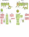
Molecular and pharmacological characteristics of KCa3.1, KCa2.1–3 and KCa1.1 channels. (A) Membrane topology of KCa3.1/KCa2.1–3 and KCa1.1 channels. Left: schematic illustration of a single KCa3.1/KCa2.1–3 subunit with six transmembrane domains (1–6) and a pore loop between S5 and S6. Ca2+ sensitivity is conferred by constitutively bound calmodulin (CaM) to the intracellular c-terminus. Right: illustration of a single α-subunit of KCa1.1 with seven transmembrane domains (0–6) and an associated β-subunit with two transmembrane domains. The extremely long intracellular c-terminus contains additional hydrophobic segments (7–10) and the so-called Ca2+ bowl, conferring Ca2+ sensitivity to gather with the hydrophobic segments 7 and 8. (B) Pharmacology of KCa3.1, KCa2.1–3 and KCa1.1 channels. Left: blockers and openers of KCa3.1 and KCa2.1–3 channels. Note that the openers have a higher affinity to KCa3.1 over KCa2.1–3 channels. Right: blockers and openers of the KCa1.1 channel. (C) KCa3.1 model (side view) based on the crystal structure of the bacterial KcsA channel (Doyle et al., 1998). Only two of the four subunits are shown to have a better view on the selectivity filter with the glycine (Gly)/tyrosine (Tyr)/glycine signature motive (in green) for a K+-selective and the cavity of the channel. The hydrophobic residues of threonine (Thr250) and valine (Val275) are lining the water filled of the cavity and are required for TRAM-34 and arachidonic acid (AA) binding just below the selectivity filter. 1-EBIO, 1-ethyl-2-benzimidazolinone; BMS-204352, [3S]-[+]-[5-chloro-2-methoxyphenyl]-1,3-dihydro-3-fluoro-6-[trifluoromethyl]-2H-indol-2-one; clotrimazole, 1-[(2-chlorophenyl)diphenylmethyl]-1H-imidazole; DC-EBIO, 5,6-dichloro-1-ethyl-1,3-dihydro-2H-benzimidazole-2-one; DHS-1, dehydrosoyasaponin-1; KCa1.1, large-conductance Ca2+-activated K+ channel; KCa2.3, small-conductance Ca2+-activated K+ channel subtype 3; KCa3.1, intermediate-conductance Ca2+-activated K+ channel; NS11021, 1-(3,5-bis-trifluoromethyl-phenyl)-3-[4-bromo-2-(1H-tetrazol-5-yl)-phenyl]-thiourea; NS1608, (N-(3-trifluoromethyl)phenyl) N′-(2-hydroxy-5-chlorophenyl) urea; NS1619, 1,3-dihydro-1-[2-hydroxy-5-(trifluoromethyl)phenyl]-5-(trifluoromethyl)-2H-benzimidazol-2-one; NS309, 3-oxime-6,7-dichloro-1H-indole-2,3-dione; SKA-20, anthra[2,1-d]thiazol-2-amine; SKA-31, naphtho[1,2-d]thiazol-2-amine; TRAM-34, 1-[(2-chlorophenyl)diphenylmethyl]-1H-pyrazole; UCL1684, 6,12,19,20,25,26-hexahydro-5,27:13,18:21,24-trietheno-11,7-metheno-7H-dibenzo[b,m][1,5,12,16]tetraazacyclotricosine-5,13-diium.
Figure 3
Figure 3
Impact of KCa3.1/KCa2.3 deficiency on KCa currents in freshly isolated carotid artery endothelial cells (CAEC). (A) Diminished currents in CAEC from KCa3.1−/− mice (KCa3.1−/−) and almost complete suppression of KCa currents in doxycycline (Dox)-administered KCa3.1−/−/KCa2.3T/T mice; wt indicates wild-type control (wt). (B) Deficiency of KCa3.1/KCa2.3 impairs acetylcholine (ACh)-induced vasodilation in murine carotid artery. Representative traces show EDHF-type vasodilation (in the presence of L-NNA and indomethacin) in response to increasing concentrations of ACh (−9 to −5 = logM [ACh]) and to the nitric oxide donor sodium nitroprusside (SNP, 10 µmol·L−1) in wt carotid artery (left) and in carotid artery from Dox-administered KCa3.1−/−/KCa2.3T/T (+Dox) mice (right). Carotid arteries were pre-constricted with phenylephrine (PE, 1 µmol·L−1). EDHF, endothelium-derived hyperpolarizing factor; KCa2.3, small-conductance Ca2+-activated K+ channel subtype 3; KCa3.1, intermediate-conductance Ca2+-activated K+ channel.
Figure 4
Figure 4
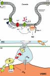
Hypothetical compartmentation of KCa3.1 and KCa2.3 channels in the endothelium. (A) Hypothetical co-localization of the small-conductance Ca2+-activated KCa channel subtype 3 (KCa2.3) together with the endothelial nitric oxide synthase (eNOS) and G protein-coupled receptor (GPCR) in caveolae [based on biochemical evidence (Weston et al., 2005)]. Note that the intermediate-conductance KCa channel (KCa3.1) is located in the less buoyant membrane fractions together with the calcium-sensing receptor (CaR). Other abbreviations: Cav-1, caveolin-1 (hairpin-like and structure-giving protein of caveolae); DAG, diacylglycerol, PLC, phospholipase C. Note that the intermediate-conductance KCa (KCa3.1) is located in the less buoyant membrane fractions. (B) KCa3.1 localization in endothelial projections facing smooth muscle cells [based on immunohistochemical evidence (Sandow et al., 2006; Dora et al., 2008; Ledoux et al., 2008b)]. Possible activation of KCa3.1 by so-called Ca2+-pulsars in response to agonist stimulation (based on high-resolution calcium-imaging experiments in arterial preparations (Ledoux et al., 2008b). EC, endothelial cell; Kir, inwardly rectifying K+ channel; meGJ, myo-endothelial gap-junction; VSMC, vascular smooth muscle cell.

Similar articles

Cited by

References

    1. Absi M, Burnham MP, Weston AH, Harno E, Rogers M, Edwards G. Effects of methyl beta-cyclodextrin on EDHF responses in pig and rat arteries; association between SK(Ca) channels and caveolin-rich domains. Br J Pharmacol. 2007;151:332–340. - PMC - PubMed
    1. Ai D, Fu Y, Guo D, Tanaka H, Wang N, Tang C, et al. Angiotensin II up-regulates soluble epoxide hydrolase in vascular endothelium in vitro and in vivo. Proc Natl Acad Sci USA. 2007;104:9018–9023. - PMC - PubMed
    1. Alexander SP, Mathie A, Peters JA. Guide to Receptors and Channels (GRAC), 3rd edition. Br J Pharmacol. 2008;153(Suppl. 2):S1–S209. - PMC - PubMed
    1. Angulo J, Cuevas P, Fernandez A, Gabancho S, Allona A, Martin-Morales A, et al. Diabetes impairs endothelium-dependent relaxation of human penile vascular tissues mediated by NO and EDHF. Biochem Biophys Res Commun. 2003;312:1202–1208. - PubMed
    1. Atkinson NS, Robertson GA, Ganetzky B. A component of calcium-activated potassium channels encoded by the Drosophila slo locus. Science. 1991;253:551–555. - PubMed

Publication types

MeSH terms

Substances