

Hypoxia inducible factor-1alpha correlates with vascular endothelial growth factor A and C indicating worse prognosis in clear cell renal cell carcinoma
- PMID: 19302703
- PMCID: PMC2664792
- DOI: 10.1186/1756-9966-28-40
Hypoxia inducible factor-1alpha correlates with vascular endothelial growth factor A and C indicating worse prognosis in clear cell renal cell carcinoma
Abstract
Background: The role of angiogenesis in the pathogenesis of renal cell carcinoma is well recognized, however, the influence of tumor cells in this activity has not yet been fully clarified. The aim of this study was to analyze the expression of hypoxia inducible factor-1alpha (HIF-1alpha), a regulatory factor of angiogenic switch, in comparison to vascular endothelial growth factor A and C (VEGF-A and VEGF-C), recognized to be involved in blood and lymph vessel neoangiogenesis, with potential association in the prognosis of patients with renal cell carcinoma.
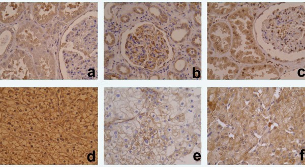
Methods: Ninety-four patients with diagnosis of clear cell renal cell carcinomas (CCRCC), all clinicopathological characteristics and overall survival were unrolled in this study. Immunohistochemicaly VEGF-A, VEGF-C, HIF-1alpha and Ki67 were detected on tumor cells and the staining was performed on tissue microarrays (TMA). The staining was evaluated as a percentage of cytoplasmic or nuclear positive tumor cells.
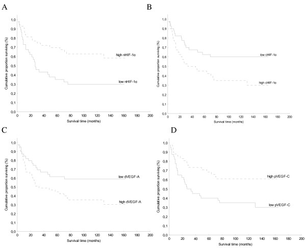
Results: Variable expression of all three proteins was confirmed. Both angiogenic factors demonstrated perimembranous or diffuse cytoplasmic staining, with diffuse pattern positively associated (p < 0.001). Nuclear HIF-1alpha expression (nHIF-1alpha) showed inverse correlation with diffuse cytoplasmic VEGF-A (p = 0.002) and VEGF-C (p = 0.053), while cytoplasmic HIF-1alpha expression (cHIF-1alpha) showed positive correlation with diffuse staining of both angiogenic factors (p < 0.001; p < 0.001, respectively). In comparison to clinicopathological characteristics, a higher nuclear grade (p = 0.006; p < 0.001, respectively), larger tumor size (p = 0.009; p = 0.015, respectively), higher stage (p = 0.023; p = 0.027, respectively) and shorter survival (p = 0.018; p = 0.024, respectively) were associated with overexpression of cHIF-1alpha and diffuse cytoplasmic VEGF-A expression. In contrary, overexpression of nHIF-1alpha was associated with better diagnostic parameters i.e. lower nuclear grade (p = 0.006), smaller tumor size (p = 0.057), and longer survival (p = 0.005).
Conclusion: Overexpression of VEGF-A and cHIF-1alpha in tumor cells highlights a more aggressive subtype of CCRCC that might have some clinical implications. The significance of nHIF-1alpha expression associated with better differentiated tumors should be further elucidated.
Figures
References
-
- Folkman J. Tumor angiogenesis: therapeutic implications. N Engl J Med. 1971;285:1182–6. - PubMed
-
- Gunningham SP, Currie MJ, Han C, Turner K, Scott PA, Robinson BA, Harris AL, Fox SB. Vascular endothelial growth factor-B and vascular endothelial growth factor-C expression in renal cell carcinomas: regulation by the von Hippel-Lindau gene and hypoxia. Cancer Res. 2001;61:3206–11. - PubMed
-
- Eble JN, Sauter G, Epstein JI, Sesterhenn IA. WHO Classification of Tumours. Pathology and Genetics of Tumours of the Urinary System and Male Genital Organs. Vol. 6. IARC Press, Lyon (France); 2004. pp. 9–87.
-
- Brieger J, Weidt EJ, Schirmacher P, Störkel S, Huber C, Decker HJ. Inverse regulation of vascular endothelial growth factor and VHL tumor suppressor gene in sporadic renal cell carcinomas is correlated with vascular growth: an in vivo study on 29 tumors. J Mol Med. 1999;77:505–10. doi: 10.1007/s001099900022. - DOI - PubMed
Publication types
MeSH terms
Substances
LinkOut - more resources
Full Text Sources
