

Cholangiocyte secretion of chemokines in experimental biliary atresia
- PMID: 19302848
- PMCID: PMC2730110
- DOI: 10.1016/j.jpedsurg.2008.07.007
Cholangiocyte secretion of chemokines in experimental biliary atresia
Abstract
Biliary atresia (BA) is a disease of the newborn that results in obstruction of the biliary tree. The cause of BA remains unknown; however, recent studies using the murine model of biliary atresia have found that rotavirus infection of the biliary epithelial cell (cholangiocyte) triggers an inflammatory response. We hypothesized that rotavirus infection of cholangiocytes results in the release of chemokines, important mediators of the host immune response.
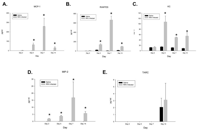
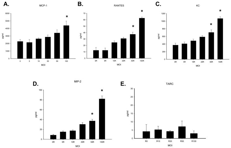
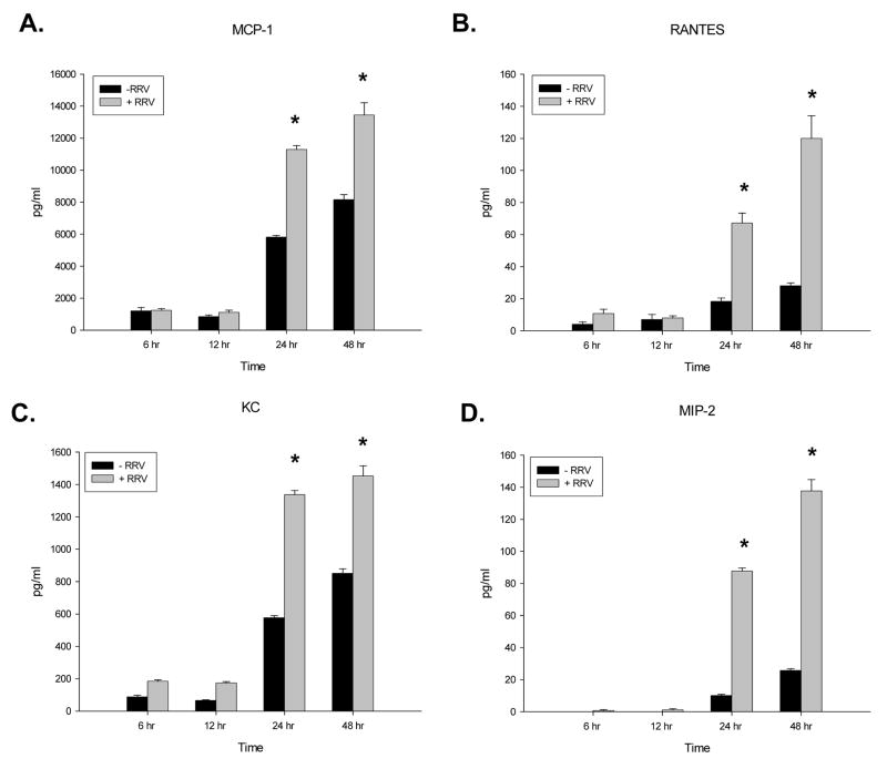
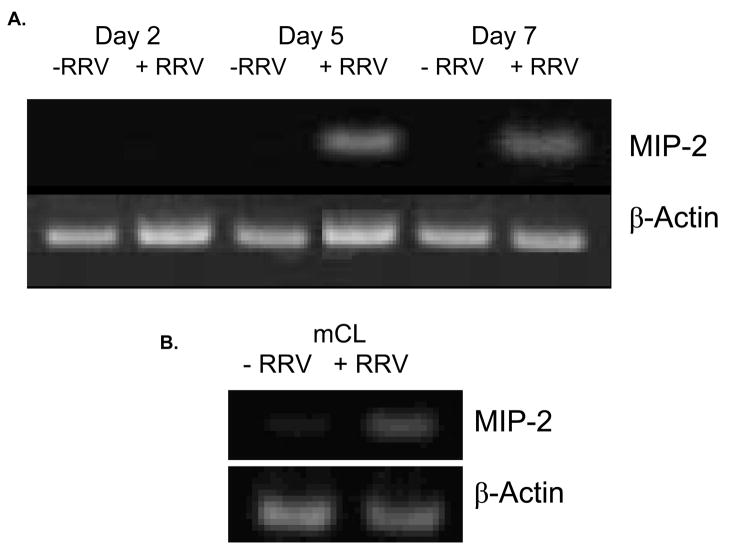
Methods: In vivo, Balb/c pups were injected with rhesus rotavirus (RRV) or saline, and, their extrahepatic bile ducts were microdissected 2, 5, 7, and 14 days after injection. Next, an immortalized cholangiocyte cell line (mCl) was incubated with RRV or serum-free media. Qualitative and quantitative chemokine assessment was performed using enzyme-linked immunosorbent assay, polymerase chain reaction, and immunohistochemistry.
Results: In vivo, increased levels of the chemokines macrophage inflammatory protein 2, monocyte chemotactic protein 1, KC and Regulated upon Activation, Normal T Expressed and Secreted were found in RRV-infected murine bile ducts. In vitro, infected mCl cells produced increasing amounts of these same chemokines in relation to dose and time.
Conclusion: These novel results suggest that chemokine expression by RRV-infected cholangiocytes may trigger a host inflammatory process that causes bile duct obstruction. Understanding how viral infection initiates this response may shed light on the pathogenesis of biliary atresia.
Figures

 ) were observed in liver samples harvested from mice on post infection day 2 and day 7. Large arrow (
 ) indicates portal vein.
References
-
- Balistreri WF, Grand R, Hoofnagle JH, Suchy FJ, Ryckman FC, Perlmutter DH, et al. Biliary atresia: current concepts and research directions. Summary of a symposium. Hepatology. 1996;23(6):1682–92. - PubMed
-
- Riepenhoff-Talty M, Gouvea V, Evans MJ, Svensson L, Hoffenberg E, Sokol RJ, et al. Detection of group C rotavirus in infants with extrahepatic biliary atresia. J Infect Dis. 1996;174(1):8–15. - PubMed
-
- Domiati-Saad R, Dawson DB, Margraf LR, Finegold MJ, Weinberg AG, Rogers BB. Cytomegalovirus and human herpesvirus 6, but not human papillomavirus, are present in neonatal giant cell hepatitis and extrahepatic biliary atresia. Pediatr Dev Pathol. 2000;3(4):367–73. - PubMed
-
- Drut R, Drut RM, Gomez MA, Cueto Rua E, Lojo MM. Presence of human papillomavirus in extrahepatic biliary atresia. J Pediatr Gastroenterol Nutr. 1998;27(5):530–5. - PubMed
-
- Fjaer RB, Bruu AL, Nordbo SA. Extrahepatic bile duct atresia and viral involvement. Pediatr Transplant. 2005;9(1):68–73. - PubMed
MeSH terms
Substances
Grants and funding
LinkOut - more resources
Full Text Sources
Research Materials
