

Hepatic stellate cells promote hepatocyte engraftment in rat liver after prostaglandin-endoperoxide synthase inhibition
- PMID: 19303017
- PMCID: PMC2693465
- DOI: 10.1053/j.gastro.2009.03.003
Hepatic stellate cells promote hepatocyte engraftment in rat liver after prostaglandin-endoperoxide synthase inhibition
Abstract
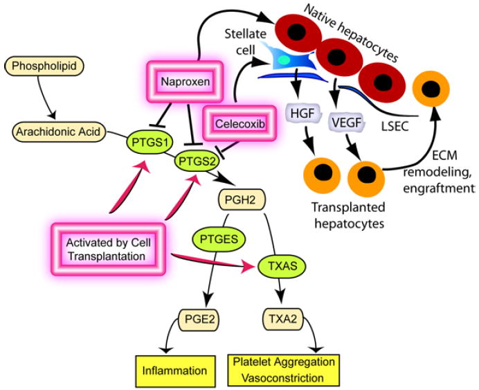
Background & aims: Hepatic inflammation occurs immediately after cells are transplanted to the liver, but the mechanisms that underlie this process are not fully defined. We examined cyclooxygenase pathways that mediate hepatic inflammation through synthesis of prostaglandins, prostacyclins, thromboxanes, and other prostanoids following transplantation of hepatocytes.
Methods: We transplanted F344 rat hepatocytes into syngeneic dipeptidyl peptidase IV-deficient F344 rats. Changes in cyclooxygenase pathways were analyzed, and specific pathways were blocked pharmacologically; the effects on cell engraftment and native liver cells were determined.
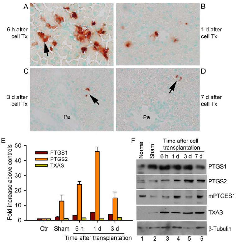
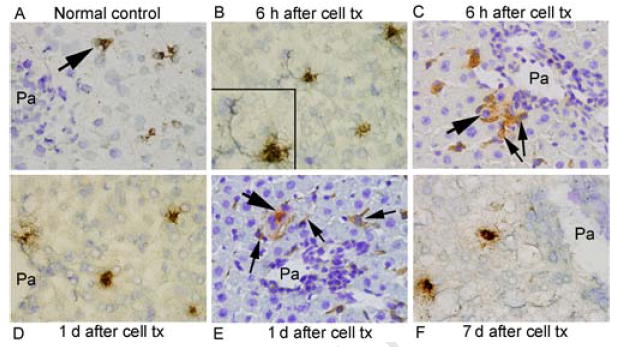
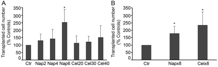
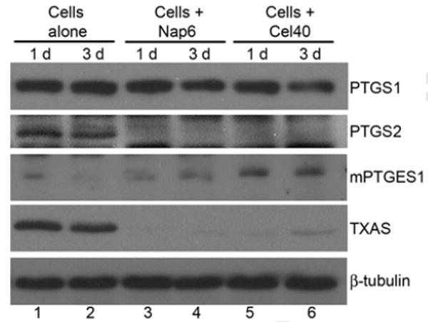
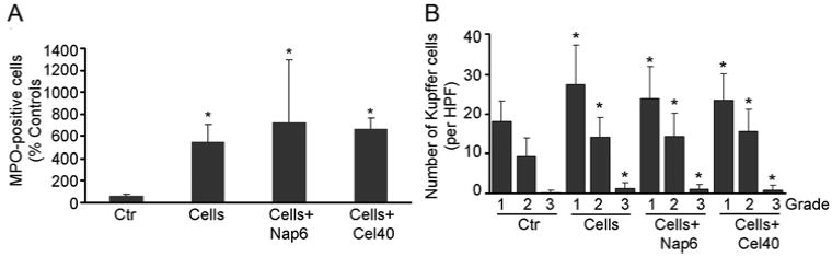
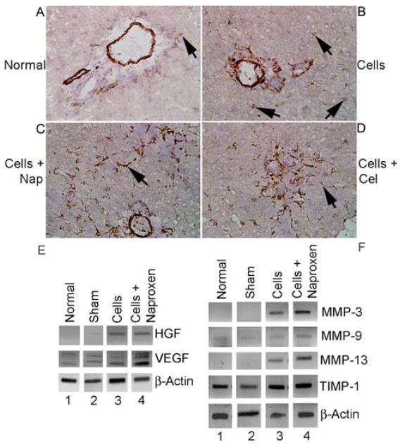
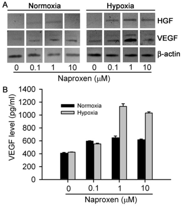
Results: Transplantation of hepatocytes induced hepatic expression of prostaglandin-endoperoxide synthases 1 and 2, which catalyze production of prostaglandin H2, as well as the downstream factor thromboxane synthase, which produces thromboxane A2 (a regulator of vascular and platelet responses in inflammation). Transplanted hepatocytes were in proximity with liver cells that expressed prostaglandin-endoperoxide synthases. The number of engrafted hepatocytes increased in rats given naproxen or celecoxib before transplantation but not in rats given furegrelate (an inhibitor of thromboxane synthase) or clopodigrel (an antiplatelet drug). Naproxen and celecoxib did not prevent hepatic ischemia or activation of neutrophils, Kupffer cells, or inflammatory cytokines, but they did induce hepatic stellate cells to express cytoprotective genes, vascular endothelial growth factor and hepatocyte growth factor, and matrix-type metalloproteinases and tissue inhibitor of metalloproteinase-1, which regulate hepatic remodeling.
Conclusions: Activation of cyclooxygenase pathways interferes with engraftment of transplanted hepatocytes in the liver. Pharmacologic blockade of prostaglandin-endoperoxide synthases stimulated hepatic stellate cells and improved cell engraftment.
Conflict of interest statement
No conflicts of interest exist
Figures
Similar articles
-
Hepatocyte transplantation activates hepatic stellate cells with beneficial modulation of cell engraftment in the rat.Hepatology. 2005 Nov;42(5):1072-81. doi: 10.1002/hep.20889. Hepatology. 2005. PMID: 16250034
-
Endothelin-1 receptor A blocker darusentan decreases hepatic changes and improves liver repopulation after cell transplantation in rats.Hepatology. 2014 Mar;59(3):1107-17. doi: 10.1002/hep.26766. Epub 2014 Jan 16. Hepatology. 2014. PMID: 24114775 Free PMC article.
-
Nonselective inhibition of prostaglandin-endoperoxide synthases by naproxen ameliorates acute or chronic liver injury in animals.Exp Mol Pathol. 2014 Feb;96(1):27-35. doi: 10.1016/j.yexmp.2013.10.017. Epub 2013 Nov 10. Exp Mol Pathol. 2014. PMID: 24220607 Free PMC article.
-
Cooperation of liver cells in health and disease.Adv Anat Embryol Cell Biol. 2001;161:III-XIII, 1-151. doi: 10.1007/978-3-642-56553-3. Adv Anat Embryol Cell Biol. 2001. PMID: 11729749 Review.
-
Celecoxib, a selective cyclooxygenase-2 inhibitor for the treatment of rheumatoid arthritis and osteoarthritis.Clin Ther. 1999 Sep;21(9):1497-513; discussion 1427-8. doi: 10.1016/s0149-2918(00)80005-3. Clin Ther. 1999. PMID: 10509845 Review.
Cited by
-
Cell therapy in end-stage liver disease: replace and remodel.Stem Cell Res Ther. 2023 May 25;14(1):141. doi: 10.1186/s13287-023-03370-z. Stem Cell Res Ther. 2023. PMID: 37231461 Free PMC article. Review.
-
Efficient Reconstitution of Hepatic Microvasculature by Endothelin Receptor Antagonism in Liver Sinusoidal Endothelial Cells.Hum Gene Ther. 2019 Mar;30(3):365-377. doi: 10.1089/hum.2018.166. Epub 2018 Dec 18. Hum Gene Ther. 2019. PMID: 30266073 Free PMC article.
-
Alternative Cell Sources for Liver Parenchyma Repopulation: Where Do We Stand?Cells. 2020 Feb 28;9(3):566. doi: 10.3390/cells9030566. Cells. 2020. PMID: 32121068 Free PMC article. Review.
-
Cell therapy to remove excess copper in Wilson's disease.Ann N Y Acad Sci. 2014 May;1315(1):70-80. doi: 10.1111/nyas.12450. Ann N Y Acad Sci. 2014. PMID: 24820353 Free PMC article. Review.
-
Human Liver Progenitor Cells for Liver Repair.Cell Med. 2013 Apr 29;5(1):1-16. doi: 10.3727/215517913X666459. eCollection 2013 Aug 10. Cell Med. 2013. PMID: 26858860 Free PMC article. Review.
References
-
- Joseph B, Malhi H, Bhargava KK, Palestro CJ, McCuskey RS, Gupta S. Kupffer cells participate in early clearance of syngeneic hepatocytes transplanted in the rat liver. Gastroenterology. 2002;123:1677–1685. - PubMed
-
- Joseph B, Kumaran V, Berishvili E, Bhargava KK, Palestro CJ, Gupta S. Monocrotaline promotes transplanted cell engraftment and advances liver repopulation in rats via liver conditioning. Hepatology. 2006;44:1411–1420. - PubMed
-
- Gupta S, Rajvanshi P, Sokhi RP, Slehria S, Yam A, Kerr A, Novikoff PM. Entry and integration of transplanted hepatocytes in liver plates occur by disruption of hepatic sinusoidal endothelium. Hepatology. 1999;29:509–519. - PubMed
-
- Sokhi RP, Rajvanshi P, Gupta S. Transplanted reporter cells help in defining onset of hepatocyte proliferation during the life of F344 rats. Am J Physiol Gastroint Liver Physiol. 2000;279:G631–G640. - PubMed
-
- Wu YM, Joseph B, Gupta S. Immunosuppression using the mTOR inhibition mechanism affects replacement of the rat liver with transplanted cells. Hepatology. 2006;44:410–9. - PubMed
Publication types
MeSH terms
Substances
Grants and funding
LinkOut - more resources
Full Text Sources
