. 2009 Jun 15;439(1-2):71-8.
doi: 10.1016/j.gene.2009.02.022. Epub 2009 Mar 18.

Splice variants of the human ZC3H14 gene generate multiple isoforms of a zinc finger polyadenosine RNA binding protein

Affiliations

Splice variants of the human ZC3H14 gene generate multiple isoforms of a zinc finger polyadenosine RNA binding protein

Sara W Leung et al. Gene. .

Abstract

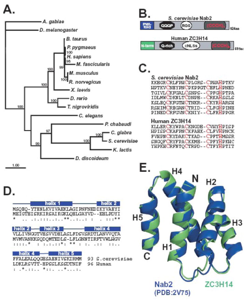
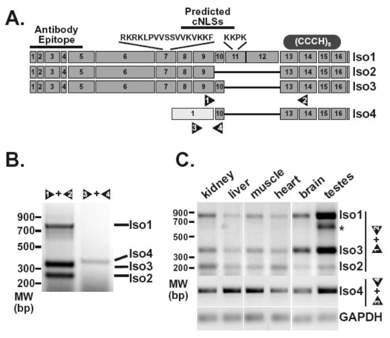
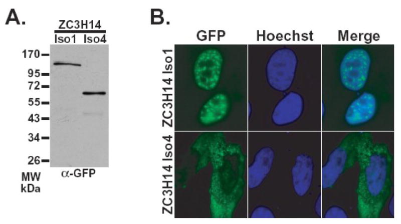
The human ZC3H14 gene encodes an evolutionarily conserved Cys(3)His zinc finger protein that binds specifically to polyadenosine RNA and is thus postulated to modulate post-transcriptional gene expression. Expressed sequence tag (EST) data predicts multiple splice variants of both human and mouse ZC3H14. Analysis of ZC3H14 expression in both human cell lines and mouse tissues confirms the presence of multiple alternatively spliced transcripts. Although all of these transcripts encode protein isoforms that contain the conserved C-terminal zinc finger domain, suggesting that they could all bind to polyadenosine RNA, they differ in other functionally important domains. Most of the alternative transcripts encode closely related proteins (termed isoforms 1, 2, 3, and 3 short) that differ primarily in the inclusion of three small exons, 9, 10, and 11, resulting in predicted protein isoforms ranging from 82 to 64 kDa. Each of these closely related isoforms contains predicted classical nuclear localization signals (cNLS) within exons 7 and 11. Consistent with the presence of these putative nuclear targeting signals, these ZC3H14 isoforms are all localized to the nucleus. In contrast, an additional transcript encodes a smaller protein (34 kDa) with an alternative first exon (isoform 4). Consistent with the absence of the predicted cNLS motifs located in exons 7 and 11, ZC3H14 isoform 4 is localized to the cytoplasm. Both EST data and experimental data suggest that this variant is enriched in testes and brain. Using an antibody that detects endogenous ZC3H14 isoforms 1-3 reveals localization of these isoforms to nuclear speckles. These speckles co-localize with the splicing factor, SC35, suggesting a role for nuclear ZC3H14 in mRNA processing. Taken together, these results demonstrate that multiple transcripts encoding several ZC3H14 isoforms exist in vivo. Both nuclear and cytoplasmic ZC3H14 isoforms could have distinct effects on gene expression mediated by the common Cys(3)His zinc finger polyadenosine RNA binding domain.


Figures

Figure 1
Figure 1. ZC3H14 is a conserved tandem zinc finger protein
A. A Bayesian phylogenetic tree of proteins related to human ZC3H14 is shown. Nodes with posterior probabilities larger than 0.7 are indicated in the figure as percentages. The accession numbers corresponding to the sequences employed for the tree reconstruction are provided in Table I. B. The similarity in the arrangement of the domains within S. cerevisiae Nab2 and human ZC3H14 is illustrated. Both proteins contain N-terminal domains that assume (S. cerevisiae) or are predicted to assume (human) a proline-tryptophan-isoleucine (PWI-fold) followed by a glutamine-rich domain (QQQP) a nuclear targeting signal (RGG or cNLS) and finally tandem Cys3His (CCCH) zinc finger motifs. Human ZC3H14 has five Cys3His (CCCH) zinc fingers while Nab2 has seven. C. Alignment of the individual C-terminal zinc finger domains of S. cerevisiae Nab2 and human ZC3H14 illustrates the similarity in spacing between the cysteine and histidine residues. D. An alignment of the N-terminal domains of Nab2 (amino acids 1-93) and ZC3H14 (amino acids 1-96) used to generate the model in panel E is shown. Identical residues are indicated by the asterisks, very similar residues by the double dots, and similar residues by single dots. E. A homology model of the N-terminal domain (amino acids 1-96) of human ZC3H14 (green) overlaid with the N-terminal domain of S. cerevisiae Nab2 (PDB accession code 2V75; blue). Homology modeling was carried out as described in Materials and Methods. Molecular images were generated using PyMol (http://pymol.sourceforge.net).
Figure 2
Figure 2. ZC3H14splice variants can be detectedin vivo
A. The diagram depicts exons predicted for the ZC3H14 splice variants encoding ZC3H14 isoform 1 (Iso1), isoform 2 (Iso2), isoform 3 (Iso3), and isoform 4 (Iso4). The sequences and approximate positions of predicted classical NLS motifs (cNLSs) are indicated at the top of the diagram. The location of the antibody epitope used to generate the polyclonal ZC3H14 antibody is also indicated as is the approximate location of the tandem Cys3His zinc finger domain (CCCH). Positions of primer pairs (1 and 2, 3 and 4) used to detect the splice variants are indicated at the bottom of the diagram. B. RT-PCR reactions (30 cycles) were performed on RNA isolated from HEK293 cells as described in Materials and Methods. Transcripts corresponding to ZC3H14 isoforms 1, 2, and 3 (Iso1, Iso2, Iso3) were detected using primer pair 1 and 2. Primer pair 3 and 4 was used to amplify the transcript corresponding to ZC3H14 isoform 4 (Iso4). Positions of the products corresponding to each splice variant are indicated. An additional band identified in the testes sample is indicated by the asterisk. This band likely reflects a ZC3H14 splice variant with additional alternative splicing of exons 10, 11, and 12. C. RT-PCR reactions were performed on RNA isolated from the mouse tissues indicated (kidney, liver, muscle, heart, brain, and testes). Isoform 1, 2 and 3 transcripts (Iso1, Iso2, Iso3) were detected using mouse primer pair 1 and 2 (see Figure 2A) with 30 cycles of amplification while the isoform 4 transcript (Iso4) was detected using mouse primer pair 3 and 4 with 60 cycles of amplification. The GAPDH transcript was amplified as a control for each tissue sample.
Figure 3
Figure 3. The ZC3H14 gene encodes both nuclear and cytoplasmic isoforms
Plasmids encoding either ZC3H14 isoform 1-GFP or isoform 4-GFP were transiently transfected into HeLa cells. A. GFP fusion proteins were detected by immunoblotting cell lysates with anti-GFP antibody (Seedorf et al., 1999). B. Transfected HeLa cells were fixed and the localization of GFP fusion proteins was examined by directed GFP fluorescence microscopy (GFP) (green). Hoechst dye (blue) was used to stain DNA and indicate the position of the nucleus. A merged image is also shown.
Figure 4
Figure 4. Endogenous nuclear ZC3H14 colocalizes with nuclear speckles
A. Lysates from HeLa cells expressing either GFP or ZC3H14 isoform 1-GFP (Iso1-GFP) fusion protein were probed with polyclonal anti-ZC3H14 (α-ZC3H14) or anti-GFP (α-GFP) antibody. B. The localization of endogenous ZC3H14 (isoforms 1-3) in HeLa cells was determined by indirect immunofluorescence using the polyclonal anti-ZC3H14 antibody. Hoechst dye was used to stain DNA and indicate the position of the nucleus. The merge reveals that endogenous ZC3H14 (isoforms 1-3) is located in foci within the nucleus. A corresponding Phase image is also shown. C. ZC3H14 localizes to nuclear speckles in HeLa cells. ZC3H14 was detected with anti-ZC3H14 antibody (green) and nuclear speckles were labeled with anti-SC-35 antibody (red). Colocalization (yellow) is indicated in the merge panel. To assess whether ZC3H14 shows dynamics typical for nuclear speckle proteins, ZC3H14 was localized either prior to (-actinomycinD) or following (+actinomycinD) treatment with actinomycinD for 4 hr. As a control, the –actinomycinD sample was treated with DMSO, which is the solvent for actinomycinD. The merge image shows that nuclear ZC3H14 colocalizes (yellow) with nuclear speckles under all conditions examined.

References

    1. Adam SA, Nakagawa T, Swanson MS, Woodruff TK, Dreyfuss G. mRNA polyadenylate-binding protein: gene isolation and sequencing and identification of a ribonucleoprotein consensus sequence. Mol Cell Biol. 1986;6:2932–43. - PMC - PubMed
    1. Aitchison JD, Blobel G, Rout MP. Kap104p: A karyopherin involved in the nuclear transport of messenger RNA binding proteins. Science. 1996;274:624–627. - PubMed
    1. Amrani N, Minet M, Le Gouar M, Lacroute F, Wyers F. Yeast Pab1 interacts with Rna15 and participates in the control of the poly(A) tail length in vitro. Mol Cell Biol. 1997;17:3694–3701. - PMC - PubMed
    1. Anderson JT, Wilson SM, Datar KV, Swanson MS. NAB2: a yeast nuclear polyadenylated RNA-binding protein essential for cell viability. Mol Cell Biol. 1993;13:2730–2741. - PMC - PubMed
    1. Apponi LH, Kelly SM, Harreman MT, Lehner AN, Corbett AH, Valentini SR. An Interaction between Two RNA Binding Proteins, Nab2 and Pub1, Links mRNA Processing/Export and mRNA Stability. Mol Cell Biol. 2007 - PMC - PubMed

Publication types

MeSH terms