. 2009 Mar 20;30(3):408-20.
doi: 10.1016/j.immuni.2009.01.010.

The lymphotoxin LTalpha(1)beta(2) controls postnatal and adult spleen marginal sinus vascular structure and function

Affiliations

The lymphotoxin LTalpha(1)beta(2) controls postnatal and adult spleen marginal sinus vascular structure and function

Carlene L Zindl et al. Immunity. .

Abstract

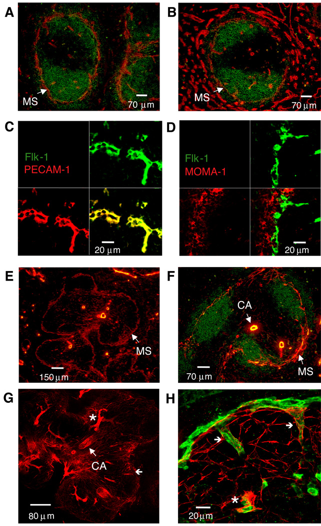
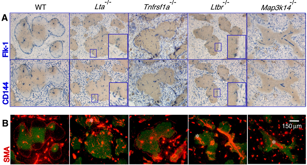
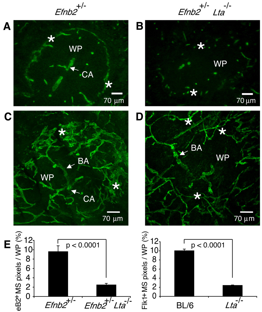
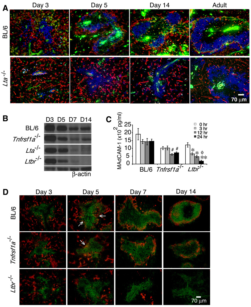
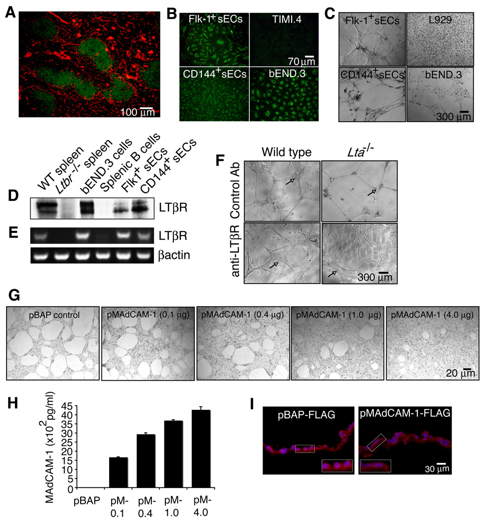
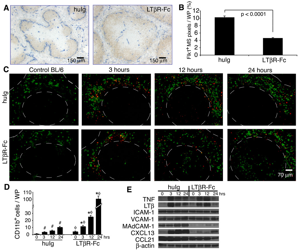
The lymphotoxin LTalpha(1)beta(2) supports the development and maintenance of several aspects of spleen structure, but its significance for marginal sinus (MS) vascular organization is unclear. We showed here that, in early postnatal lymphotoxin-deficient mice, the developing Flk-1+ white pulp vessels failed to organize or upregulate MAdCAM-1, leading to altered spatial rearrangement of both the white pulp endothelial cells and the smooth muscle actin-expressing cells. In vitro, MAdCAM-1 directed the reorganization of LTbeta receptor+ endothelial cells grown on Matrigel. LTalpha(1)beta(2) also regulated the maintenance of both MAdCAM-1 expression and mature MS structure in adult mice, contributing importantly to normal trafficking of CD11b+ cells in response to bacterial antigens. Together, our studies demonstrate that LTalpha(1)beta(2) and LTbeta receptor signals control proper development and maintenance of the mature MS structure and implicate MAdCAM-1 in the structuring of the MS endothelial cells that is important for the movement of immune cells within the spleen.


Figures

Figure 1
Figure 1. Flk-1 marks marginal sinus smooth muscle-associated endothelial cells in the mouse spleen
Ten µm thick frozen sections from C57BL/6 spleens (n>10) were stained with anti-B220 (green) and either (A) anti-Flk-1 (red) or (B) anti-CD144 (VE-cadherin) (red) (magnification 200×). In panels C and D, staining was with anti-Flk-1 (green) and either (C) anti-PECAM-1 (red) or (D) anti-MOMA-1 (red). For panels E and F, spleen sections were cut at 20µm and stained with (E) anti-SMA alone (red) (magnification 100×) or together with (F) anti-B220 (green) (magnification 200×). For panels G and H, staining was with (G) anti-SMA alone (red) (magnification 100×) or together with (H) anti-Flk-1 (green) (magnification 630×). For panels C, D, G, and H, confocal microscopy was used to compile a series of Z-stack images to reconstruct a 6 µm thick section. CA, central arteriole; *, central arteriole branching vessel; (←), MS connecting vessels. Similar data were obtained in two additional experiments.
Figure 2
Figure 2. LTβR signaling supports the organization of endothelial and smooth muscle cells in the marginal sinus
Twenty µm thick frozen sections from spleens (n≥5) of the indicated mouse strains were stained with (A) anti-Thy1.2 (brown) and either anti-Flk-1 (blue, top panels) or anti-CD144 (VE-cadherin) (blue, bottom panels), and (B) anti-Thy1.2 (green) and anti-SMA (red) (magnification 200×). Inset images show magnified areas highlighting the Flk-1+ and CD144+ structures at the periphery of white pulp areas in the gene targeted mice.
Figure 3
Figure 3. Disturbed MS endothelial cell network in Lta−/− mice
Spleens of Efnb2+/− (A, B) and Efnb2+/− Lta−/− (C, D) mice were cut to yield (A, C) 10 µm or (B, D) 120 µm sections and stained with anti-β-galactosidase (green) to visualize ephrinB2 expression via the expression of the in frame knock in of the LacZ gene. Confocal images were compiled to yield a 50–60 µm section of spleen, permitting analysis of the 3-D structure of a region of a single white pulp nodule (magnification 200×). Similar data were obtained by analyzing spleens from 5 additional pairs of mice. (E) The percentage of ephrinB2+ and Flk-1+ MS structures per WP area was quantified from 10 µm sections using a 20× objective. Graphs show mean + SD (n≥40 WP nodules using ≥5 spleens per strain). Data were compiled from 5 independent experiments.
Figure 4
Figure 4. Lymphotoxin controls organization of Flk-1+ vessels and expression of MAdCAM-1 during neonatal development
(A) Frozen sections of spleens from C57BL/6 and Lta−/− mice harvested at the indicated ages (n≥5 per strain per time point) were stained with anti-Flk-1 (red), anti-SMA (green), and anti-B220 (blue) (magnification 200×). Expression of MAdCAM-1 protein was analyzed by (B) immunoblotting and (C) ELISA from extracts of whole spleens (n=10 spleens per strain per time point) of C57BL/6, Lta−/−, Ltbr−/− and Tnfrsf1a−/− mice. (D) Spleen sections from the indicated mouse strains (n=10 spleens per strain per time point) harvested on the indicated postnatal days were stained with anti-MAdCAM-1 (red) and anti-B220 (green). #p <0.01 comparing C57BL/6 and Tnfrsf1a−/− mice, *p <0.005 and **p <0.001 for comparisons between C57BL/6 and Ltbr−/− mice, and ϕp <0.001 for comparison between Tnfrsf1a−/− and Ltbr−/− mice.
Figure 5
Figure 5. Endothelial cells express the LTβR and utilize MAdCAM-1 to form specialized structures on Matrigel
(A) Ten µm spleen sections from WT mice were stained with anti-LTβR (red) and anti-B220 (green) (n=3). Endothelial cells isolated from WT spleen (sECs) using either anti-Flk-1 or anti-CD144 were measured for (B) uptake of AcLDL (green) and (C) observed for formation of tube-like structures on Matrigel (n=3). sECs and the bEND.3 cell line were analyzed for expression of (D) LTβR protein and (E) RNA by immunoblotting and RT-PCR. (F) sECs isolated from WT and Lta−/− mice were plated on Matrigel containing a control GST antibody or an agonist anti-LTβR and cultured for 10 days. (G) bEND.3 cells were transfected with a control pBAP-FLAG vector (expressing bacterial alkaline phosphatase) or 0.1, 0.4, 1, and 4 µg of a pMAdCAM-1-FLAG vector. The cells were then plated on Matrigel and cultured for 24 hrs. (H) Protein extracts from transfected bEND.3 cells were prepared and an ELISA was performed to assess MAdCAM-1 protein expression. (I) Transfected bEND.3 cells grown on Anapore filters coated with Matrigel were fixed and frozen in O.C.T. and 8 µm cross-section slices were stained with anti-FLAG (red) and Hoechst (blue). Inset images show magnified areas highlighting the EC phenotype and amount of MAdCAM-1 associated with the Matrigel. Data shown are representative of 3 independent experiments.
Figure 6
Figure 6. Intact marginal sinus organization in adult mice is LT-dependent and supports the localization of immune cells within the spleen following challenge by bacterial antigens
(A) C57BL/6 adult mice were treated i.p. with either human IgG1 (huIg) or LTβR-Fc and spleens were harvested 2 wks later. 10 µm sections were stained with anti-Flk-1 (blue) and anti-B220 (brown). Similar results were obtained in 4 additional experiments. (B) The percentage of Flk-1+ MS structures per WP area was quantified from 10 µm sections using a 20× objective (n≥40 WP nodules from 3 independent experiments; graph shows mean + S.D.). (C) C57BL/6 mice were treated i.p. with either huIg or LTβR-Fc and 2 wks later were injected i.v. with 250 µg of TMR-labeled S. aureus bioparticles (red). Spleens were harvested at 3, 12 or 24 hrs after injection with the particles and sections were stained with anti-CD11b (green). Dashed lines outline WP areas. (D) The numbers of CD11b+ cells localized in the WP was quantified from ≥40 WP areas from 3 independent experiments with 3–5 mice per group. Graph shows mean + S.D. (E) Expression levels of the indicated cytokines, chemokines and adhesion molecules were analyzed by immunoblotting. Data are representative of at least 3 independent experiments with 3–5 mice per group.

Similar articles

Cited by

References

    1. Balazs M, Horvath G, Grama L, Balogh P. Phenotypic identification and development of distinct microvascular compartments in the postnatal mouse spleen. Cell Immunol. 2001;212:126–137. - PubMed
    1. Balazs M, Martin F, Zhou T, Kearney J. Blood dendritic cells interact with splenic marginal zone B cells to initiate T-independent immune responses. Immunity. 2002;17:341–352. - PubMed
    1. Balogh P, Balazs M, Czompoly T, Weih DS, Arnold HH, Weih F. Distinct roles of lymphotoxin-beta signaling and the homeodomain transcription factor Nkx2.3 in the ontogeny of endothelial compartments in spleen. Cell Tissue Res. 2007;328:473–486. - PubMed
    1. Banks TA, Rouse BT, Kerley MK, Blair PJ, Godfrey VL, Kuklin NA, Bouley DM, Thomas J, Kanangat S, Mucenski ML. Lymphotoxin-alpha-deficient mice. Effects on secondary lymphoid organ development and humoral immune responsiveness. J Immunol. 1995;155:1685–1693. - PubMed
    1. Bohnsack JF, Brown EJ. The role of the spleen in resistance to infection. Annu Rev Med. 1986;37:49–59. - PubMed

MeSH terms