Early blockade of injured primary sensory afferents reduces glial cell activation in two rat neuropathic pain models
- PMID: 19303429
- PMCID: PMC2777638
- DOI: 10.1016/j.neuroscience.2009.03.016
Early blockade of injured primary sensory afferents reduces glial cell activation in two rat neuropathic pain models
Abstract
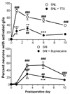
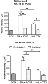
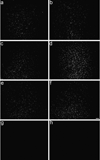
Satellite glial cells in the dorsal root ganglion (DRG), like the better-studied glia cells in the spinal cord, react to peripheral nerve injury or inflammation by activation, proliferation, and release of messengers that contribute importantly to pathological pain. It is not known how information about nerve injury or peripheral inflammation is conveyed to the satellite glial cells. Abnormal spontaneous activity of sensory neurons, observed in the very early phase of many pain models, is one plausible mechanism by which injured sensory neurons could activate neighboring satellite glial cells. We tested effects of locally inhibiting sensory neuron activity with sodium channel blockers on satellite glial cell activation in a rat spinal nerve ligation (SNL) model. SNL caused extensive satellite glial cell activation (as defined by glial fibrillary acidic protein [GFAP] immunoreactivity) which peaked on day 1 and was still observed on day 10. Perfusion of the axotomized DRG with the Na channel blocker tetrodotoxin (TTX) significantly reduced this activation at all time points. Similar findings were made with a more distal injury (spared nerve injury model), using a different sodium channel blocker (bupivacaine depot) at the injury site. Local DRG perfusion with TTX also reduced levels of nerve growth factor (NGF) in the SNL model on day 3 (when activated glia are an important source of NGF), without affecting the initial drop of NGF on day 1 (which has been attributed to loss of transport from target tissues). Local perfusion in the SNL model also significantly reduced microglia activation (OX-42 immunoreactivity) on day 3 and astrocyte activation (GFAP immunoreactivity) on day 10 in the corresponding dorsal spinal cord. The results indicate that early spontaneous activity in injured sensory neurons may play important roles in glia activation and pathological pain.
Figures


Similar articles
-
Mechanical hypersensitivity, sympathetic sprouting, and glial activation are attenuated by local injection of corticosteroid near the lumbar ganglion in a rat model of neuropathic pain.Reg Anesth Pain Med. 2011 Jan-Feb;36(1):56-62. doi: 10.1097/AAP.0b013e318203087f. Reg Anesth Pain Med. 2011. PMID: 21455091 Free PMC article.
-
Mirror-image pain is mediated by nerve growth factor produced from tumor necrosis factor alpha-activated satellite glia after peripheral nerve injury.Pain. 2014 May;155(5):906-920. doi: 10.1016/j.pain.2014.01.010. Epub 2014 Jan 18. Pain. 2014. PMID: 24447514
-
Activation of satellite glial cells in lumbar dorsal root ganglia contributes to neuropathic pain after spinal nerve ligation.Brain Res. 2012 Jan 3;1427:65-77. doi: 10.1016/j.brainres.2011.10.016. Epub 2011 Oct 14. Brain Res. 2012. PMID: 22050959
-
[Contribution of primary sensory neurons and spinal glial cells to pathomechanisms of neuropathic pain].Brain Nerve. 2008 May;60(5):483-92. Brain Nerve. 2008. PMID: 18516970 Review. Japanese.
-
Gliopathic pain: when satellite glial cells go bad.Neuroscientist. 2009 Oct;15(5):450-63. doi: 10.1177/1073858409336094. Neuroscientist. 2009. PMID: 19826169 Free PMC article. Review.
Cited by
-
Emerging importance of satellite glia in nervous system function and dysfunction.Nat Rev Neurosci. 2020 Sep;21(9):485-498. doi: 10.1038/s41583-020-0333-z. Epub 2020 Jul 22. Nat Rev Neurosci. 2020. PMID: 32699292 Free PMC article. Review.
-
Tetrodotoxin (TTX) as a therapeutic agent for pain.Mar Drugs. 2012 Feb;10(2):281-305. doi: 10.3390/md10020281. Epub 2012 Jan 31. Mar Drugs. 2012. PMID: 22412801 Free PMC article. Review.
-
John J. Bonica Award Lecture: Peripheral neuronal hyperexcitability: the "low-hanging" target for safe therapeutic strategies in neuropathic pain.Pain. 2020 Sep;161 Suppl 1(Suppl 1):S14-S26. doi: 10.1097/j.pain.0000000000001838. Pain. 2020. PMID: 33090736 Free PMC article.
-
Delayed activation of spinal microglia contributes to the maintenance of bone cancer pain in female Wistar rats via P2X7 receptor and IL-18.J Neurosci. 2015 May 20;35(20):7950-63. doi: 10.1523/JNEUROSCI.5250-14.2015. J Neurosci. 2015. PMID: 25995479 Free PMC article.
-
Microglia in Pain: Detrimental and Protective Roles in Pathogenesis and Resolution of Pain.Neuron. 2018 Dec 19;100(6):1292-1311. doi: 10.1016/j.neuron.2018.11.009. Neuron. 2018. PMID: 30571942 Free PMC article. Review.
References
-
- Biber K, Neumann H, Inoue K, Boddeke HW. Neuronal 'On' and 'Off' signals control microglia. Trends Neurosci. 2007;30:596–602. - PubMed
-
- Boucher TJ, Okuse K, Bennett DL, Munson JB, Wood JN, McMahon SB. Potent analgesic effects of GDNF in neuropathic pain states. Science. 2000;290:124–127. - PubMed
-
- Burnstock G. Physiology and pathophysiology of purinergic neurotransmission. Physiol Rev. 2007;87:659–797. - PubMed
-
- Canady KS, Ali-Osman F, Rubel EW. Extracellular potassium influences DNA and protein syntheses and glial fibrillary acidic protein expression in cultured glial cells. Glia. 1990;3:368–374. - PubMed
Publication types
MeSH terms
Substances
Grants and funding
LinkOut - more resources
Full Text Sources
Medical
Research Materials
Miscellaneous

