. 2009 Sep;35(6):802-10.
doi: 10.1016/j.burns.2008.12.013. Epub 2009 Mar 20.

Effect of ablated bronchial blood flow on survival rate and pulmonary function after burn and smoke inhalation in sheep

Affiliations

Effect of ablated bronchial blood flow on survival rate and pulmonary function after burn and smoke inhalation in sheep

Atsumori Hamahata et al. Burns. 2009 Sep.

Abstract

The bronchial circulation plays a significant role in the pathophysiological changes of burn and smoke-inhalation injury. Bronchial blood flow markedly increases immediately after inhalational injury. This study examines whether the ablation of the bronchial artery attenuates pathophysiological changes and improves survival after burn and smoke-inhalational injury in an ovine model. Acute lung injury was induced by 40% total body surface-area third-degree cutaneous burn and cotton smoke inhalation (48 breaths of cotton smoke, <40 degrees C) under deep anaesthesia. Twelve adult female sheep were divided into two groups: (1) sham (injured, non-ablated bronchial artery, n=6); (2) ablation (injured, ablated bronchial artery, n=6). Ablation of the bronchial artery was performed 72 h before the injury. The experiment was continued for 96 h. Burn and smoke-inhalation injury significantly increased regional blood flow in the bronchi. Ablation of the bronchial artery significantly reduced acute regional blood flow increases in the proximal and distal bronchi. All animals in the ablation group survived to 96 h. Four of these were successfully weaned off the ventilator. Three animals of the sham group met standardised euthanasia criteria at 60 h, while another met the criteria at 78 h. The lung wet-to-dry weight ratio, histology score and myeloperoxidase (MPO) activity were significantly increased by the insult, but ablation of the bronchial artery attenuated these changes. Burn and smoke-inhalation injury induced a significant increase in bronchial blood flow and accelerated airway obstruction, pulmonary vascular changes, pulmonary oedema and pulmonary dysfunction. Ablated bronchial circulation attenuated these pathophysiological changes.


Figures

Figure 1
Figure 1
Changes in airway blood flow after burn and smoke inhalation injury. Burn and smoke inhalation injury significantly increased the airway blood flow. Bronchial ablation didn’t reduce the changes in trachea (A). However, bronchial ablation significantly prevented the airway blood flow increases in proximal and distal bronchi (B,C). Data are expressed as mean±SE. * Significant difference vs. sham animals (p < 0.05), † Significant difference vs. Base line value (p < 0.05).
Figure 2
Figure 2
All ablation group animals survived until 96h. Four of them were succeeded of ventilation weaning. Three animals in sham group died at 60h and one of them died at 78h. Ablation group has significance difference vs. sham group in survival proportions (p=0.02).
Figure 3
Figure 3
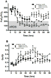
Effect of ablated bronchial blood flow on PaO2/FIO2 (A) and pulmonary shunt fraction (B). After 48h, four of ablation group’s animals was separated to ventilation weaning group (VW) and none ventilation weaning group (NVW). Data are expressed as mean±SE.
Figure 4
Figure 4
Effect of ablated bronchial artery blood flow on pulmonary arterial pressure (PAP) (A) and pulmonary vascular resistance index (PVRI) (B). After 48h, ablation group was separated to ventilation weaning group (VW) and none ventilation weaning group (NVW). Burn and smoke inhalation injury significantly increased PAP and PVRI in sham group. Ablation of bronchial artery significantly attenuated the increases of PAP at 12h, 36h and 48h and PVRI at 12h, 18h, 24h, 30h and 36h. Data are expressed as mean±SE.* Significant difference vs. sham animals (p < 0.05), † Significant difference vs. Base line value (p < 0.05).
Figure 5
Figure 5
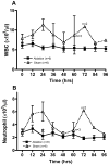
Effect of ablated bronchial artery blood flow on white blood cells counts (WBC) (A) and neutrophil counts (B). Burn and smoke inhalation injury increased WBC and neutrophil counts in sham group animals. However, ablation of bronchial artery attenuated the changes. Data are expressed as mean±SE. * Significant difference vs. sham animals (p < 0.05).
Figure 6
Figure 6
Pathological results. Each graph shows Airway obstruction score in bronchi (A), Airway obstruction score in bronchiole (B), polymorphonuclear (PMN) cells score (C) and edema formation score (D). Sham groups were separated to 60h (n=3), 78h (n=1) and 96h (n=2) following their survival time. Burn and smoke inhalation injury remarkably increased airway obstruction score in bronchia and bronchiole and PMN cells score and edema formation score. Ablated bronchial artery significantly attenuated the increase of airway abstraction score in bronchia and bronchiole and PMN cells score and edema formation score compare to sham groups at 60h. Data are expressed as mean±SE. * Significant difference vs. 60h sham animals (p < 0.05).
Figure 7
Figure 7
Effect of ablated bronchial artery blood flow on Bloodless lung wet to dry ratio (A), Myeloperoxidase in lung tissue (B). Sham groups were separated to 60h (n=3), 78h (n=1) and 96h (n=2) following their survival time. Burn and smoke inhalation injury remarkably increased lung bloodless wet to dry ratio and myeloperoxidase in lung tissue. Ablated bronchial artery significantly attenuated the increase of lung bloodless wet to dry ratio and myeloperoxidase in lung tissue compare to sham groups at 60h. Data are expressed as mean±SE. * Significant difference vs. 60h sham animals (p < 0.05).

References

    1. Saffle JR, Davis B, Williams P. Recent outcomes in the treatment of burn injury in the United States: a report from the American Burn Association Patient Registry. J Burn Care Rehabil. 1995;16(3 Pt 1):219–32. discussion 288–9. - PubMed
    1. Barrow RE, Spies M, Barrow LN, Herndon DN. Influence of demographics and inhalation injury on burn mortality in children. Burns. 2004;30(1):72–7. - PubMed
    1. Stothert JC, Jr, Ashley KD, Kramer GC, et al. Intrapulmonary distribution of bronchial blood flow after moderate smoke inhalation. J Appl Physiol. 1990;69(5):1734–9. - PubMed
    1. Linares HA. A report of 115 consecutive autopsies in burned children: 1966–80. Burns Incl Therm Inj. 1982;8(4):263–70. - PubMed
    1. Linares HA, Herndon DN, Traber DL. Sequence of morphologic events in experimental smoke inhalation. J Burn Care Rehabil. 1989;10(1):27–37. - PubMed

Publication types

MeSH terms