. 2009 Oct;16(10):982-8.
doi: 10.1016/j.phymed.2009.01.004. Epub 2009 Mar 20.

Modulation of in vitro murine B-lymphocyte response by curcumin

Affiliations

Modulation of in vitro murine B-lymphocyte response by curcumin

D Decoté-Ricardo et al. Phytomedicine. 2009 Oct.

Abstract

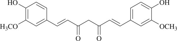
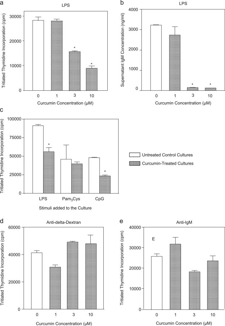
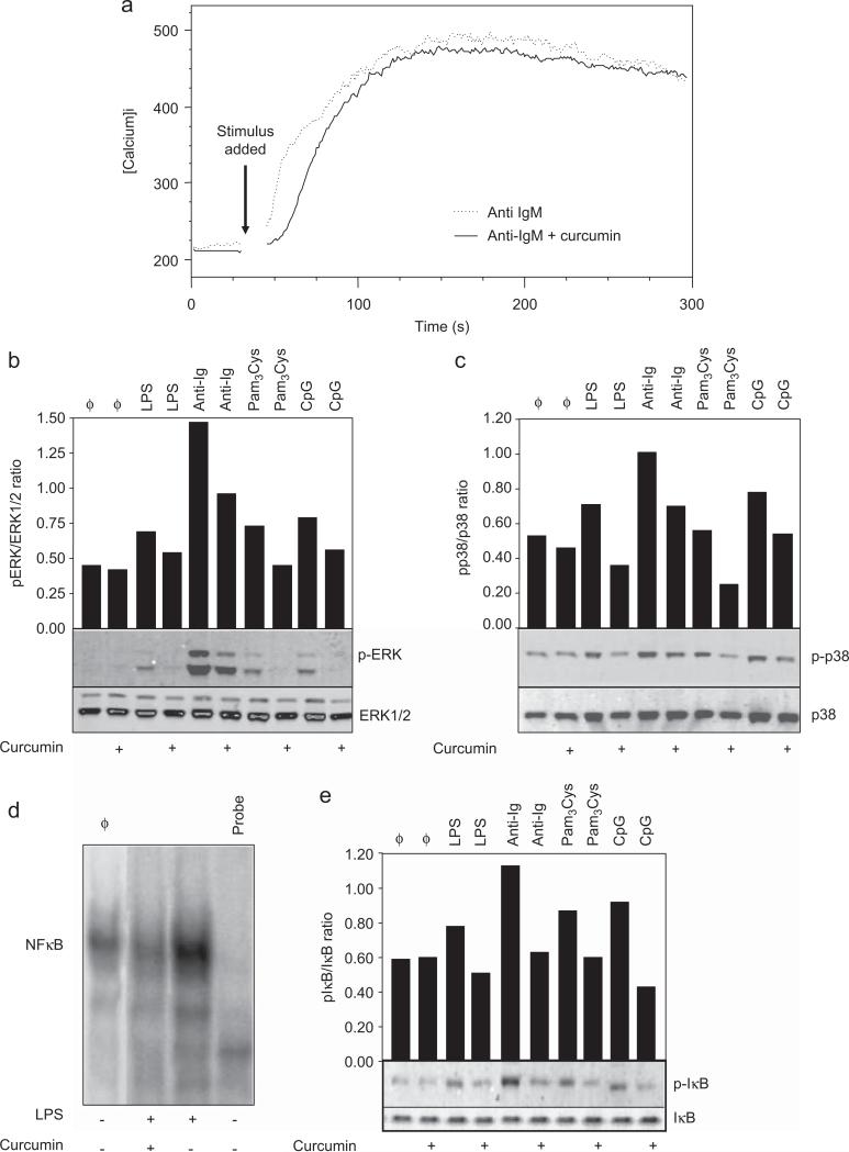
Curcumin is a phenolic natural product isolated from the rhizome of Curcuma longa (tumeric). It was previously described that curcumin had a potent anti-inflammatory effect and inhibited the proliferation of a variety of tumor cells. In the present study, we investigated the inhibitory effects of curcumin on the response of normal murine splenic B cells. Curcumin inhibited the proliferative response of purified splenic B cells from BALB/c mice stimulated with the Toll-like receptor ligands LPS and CpG oligodeoxynucleotides. LPS-induced IgM secretion was also inhibited by curcumin. The proliferative response induced by either the T-independent type 2 stimuli anti-delta-dextran or anti-IgM antibodies was relatively resistant to the effect of curcumin. We investigated the intracellular signaling events involved in the inhibitory effects of curcumin on murine B cells. Curcumin did not inhibit the increase in calcium levels induced by anti-IgM antibody. Western blotting analysis showed that curcumin inhibited TLR ligands and anti-IgM-induced phosphorylation of ERK, IkappaB and p38. Curcumin also decreased the nuclear levels of NFkappaB. Our results suggested that curcumin is an important inhibitor of signaling pathways activated upon B cell stimulation by TLR ligands. These data indicate that curcumin could be a potent pharmacological inhibitor of B cell activation.


Figures

Fig. 1
Fig. 1
Formula of curcumin.
Fig. 2
Fig. 2. Curcumin inhibits B cell proliferation and IgM production induced by TLR ligands but not by ligands to surface Ig
(a) LPS-induced proliferation. Purified B cells from BALB/c mice (1 × 10⁶/ml) were stimulated with LPS (10 μg/ml). The cultures were set in triplicate in a final volume of 200 μl and were treated simultaneously with the indicated doses of curcumin. No curcumin was added at control cultures. Proliferation was measured in cultures carried out for 48 h by the addition of tritiated thymidine (0.5 μCi per well) during the final 24 h of culture. Data are expressed as mean counts per minute (cpm) levels of triplicate cultures. Control untreated cultures showed <1200 cpm. (b) LPS-induced immunoglobulin secretion. Cultures of purified B cells (2.5 × 10⁵/ml) were set in triplicate in a final volume of 200 μl and were stimulated with LPS (10 μg/ml). Cultures were treated simultaneously with the indicated doses of curcumin. Pooled supernatants from seven days cultures were obtained and IgM levels were determined by ELISA. Control cultures had undetectable levels of IgM (<10 ng/ml). (c) TLR ligands-induced proliferation. Cultures were set as described in (a) and were stimulated with LPS (10 μg/ml), Pam3Cys (1 μg/ml) or CpG (1 μg/ml). Some cultures were also treated with curcumin (10 μM). No curcumin was added to control cultures. Proliferation was measured as described in (a). Control untreated cultures showed <1130 cpm. (d) Anti-delta-dextran-induced proliferation. B cells were stimulated with anti-delta-dextran (10 ng/ml) and curcumin was added at the indicated doses. Cultures were set and analyzed as described in (a). Control untreated cultures showed <1240 cpm. (e) Anti-IgM-induced proliferation. B cells were stimulated with anti-mouse IgM (5 μg/ml) and curcumin was added at the indicated doses. Cultures were set and analyzed as described in (a). Control untreated cultures showed <1210 cpm. Data in all figures are representative of three independent experiments. *statistically significant data (p ≤ 0.05).
Fig. 3
Fig. 3. Effect of curcumin in B cell signaling transduction
(a) Intracelular calcium levels. Purified splenic murine B cells were loaded with Indo-AM and then stained with anti-B220-FITC antibody. Baseline calcium levels were determined by flow cytometry and the cells were then stimulated with anti-IgM (10 μg/ml) and treated with curcumin (10 μM). The mean calcium levels were determined by measurement of the fluorescence ratio of Indo-1-AM (390 nm/490 nm emission) and analyzed by the FlowJo software. Data are representative of three independent experiments. (b and c) MAPK phosphorylation. Purified B cells were stimulated with LPS (10 μg/ml), CpG oligodeoxynucleotides (1 μg/ml), Pam3Cys (1 μg/ml) or anti-IgM antibody (10 μg/ml). Some cultures were simultaneously treated with curcumin (10 μM). Cells were then lysed after either a 5 min (anti-IgM-treated cells) or a 30 min (TLR ligands-treated cells) treatment and whole-cell lysates were loaded onto SDS-PAGE gels. Blot were run and probed with the following antibodies: (b) anti-phospho ERK antibody; (c) anti-phospho-p38 antibody. The same blots were then stripped and reprobed with antibodies to nonphosphorylated proteins to determine absolute protein levels. Bar graphics shows the ratio of phosphorylated and total proteins. Data are representative of five independent experiments. (d) NFκB translocation to the nucleus. Purified B cells were treated with curcumin (10 μM) and stimulated with LPS (10 μg/ml) overnight. Nuclear extracts were prepared and tested for NFκB binding activity by EMSA using a double-stranded NF-κB consensus oligonucleotide. A double-stranded mutated oligonucleotide was used to examine the specificity of NFκB binding. The specific NF-κB bands are indicated. (e) IκB phosphorylation. B cells were stimulated with LPS (10 μg/ml), CpG (1 μg/ml), Pam3Cys (1 μg/ml) or anti-IgM antibody (10 μg/ml). Curcumin was added at 10 μM where indicated. Cells were then lysed after either 5 min (anti-IgM-treated cells) or 30 min (TLR ligands-treated cells) treatment and whole-cell lysates were loaded onto SDS-PAGE gels. Blot were run and probed with anti-phospho-IκB antibody. The same blots were then stripped and reprobed with anti-IκB antibody to determine absolute protein levels. Bar graphics shows the ratio of phosphorylated and total proteins. Data shown are representative of three independent experiments.

References

    1. Aggarwal BB, Shishodia S. Molecular targets of dietary agents for prevention and therapy of cancer. Biochem. Pharmacol. 2006;71:1397–1421. - PubMed
    1. Benschop RJ, Aviszus K, Zhang X, Manser T, Cambier JC, Wysocki LJ. Activation and anergy in bone marrow B cells of a novel immunoglobulin transgenic mouse that is both hapten specific and autoreactive. Immunity. 2001;14:33–43. - PubMed
    1. Bijsterbosch MK, Meade CJ, Turner GA, Klaus GG. B lymphocyte receptors and polyphosphoinositide degradation. Cell. 1985;41:999–1006. - PubMed
    1. Brunswick M, Finkelman FD, Highet PF, Inman JK, Dintzis HM, Mond JJ. Picogram quantities of anti-Ig antibodies coupled to dextran induce B cell proliferation. J. Immunol. 1988;140:3364–3372. - PubMed
    1. Churchill M, Chadburn A, Bilinski RT, Bertagnolli MM. Inhibition of intestinal tumors by curcumin is associated with changes in the intestinal immune cell profile. J. Surg. Res. 2000;89:169–175. - PubMed

Publication types

LinkOut - more resources