. 2009 May 10;387(2):313-21.
doi: 10.1016/j.virol.2009.02.026. Epub 2009 Mar 21.

Restriction of HIV-1 by APOBEC3G is cytidine deaminase-dependent

Affiliations

Restriction of HIV-1 by APOBEC3G is cytidine deaminase-dependent

Edward P Browne et al. Virology. .

Abstract

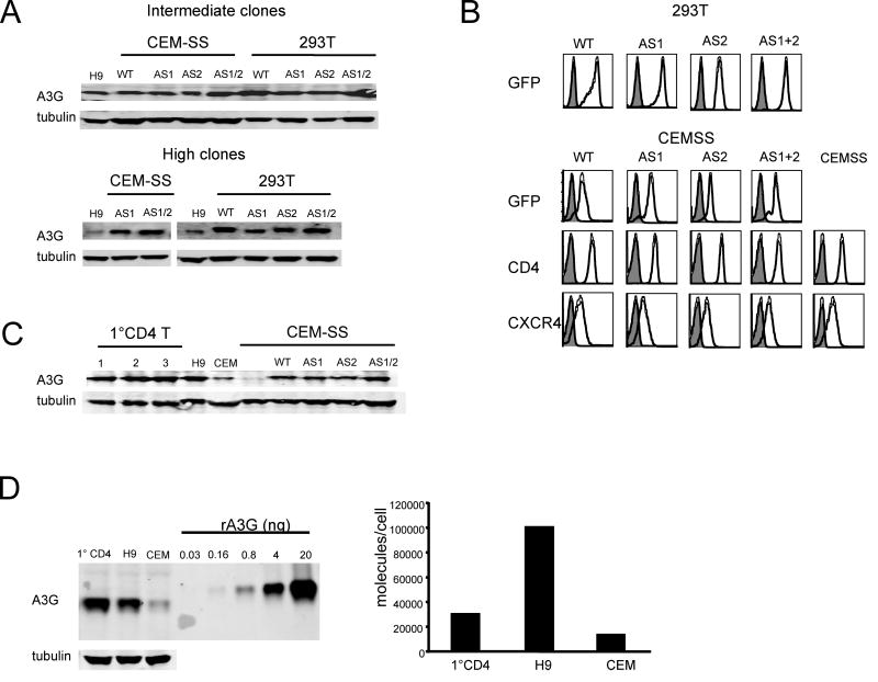
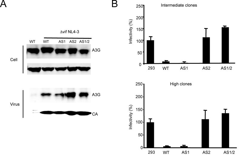
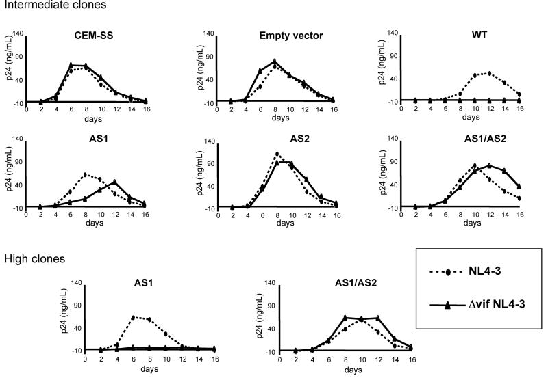
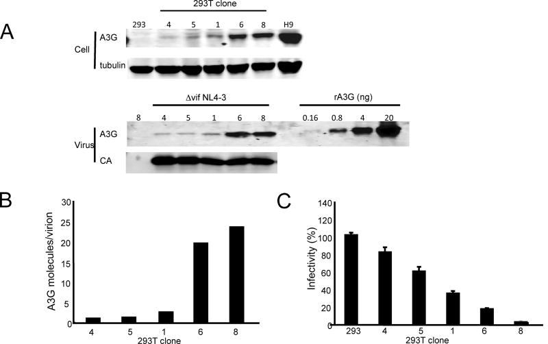
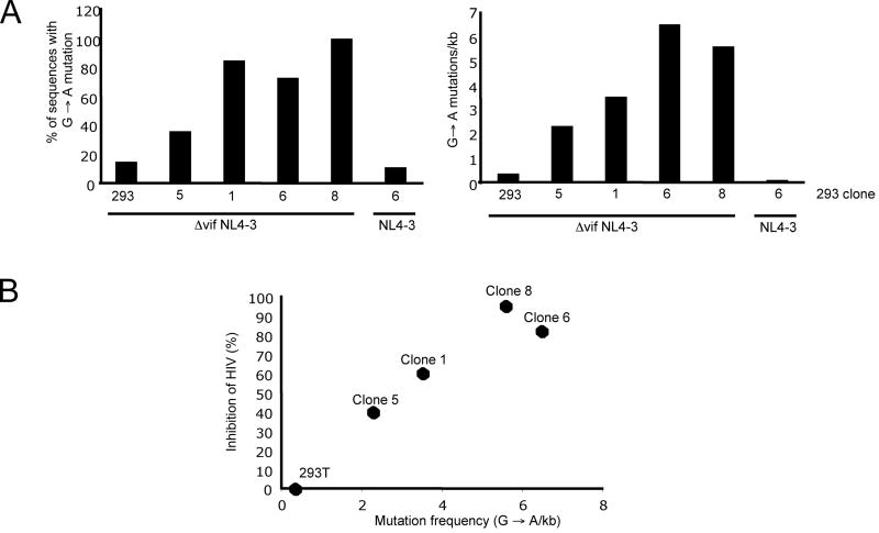
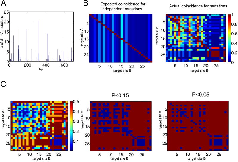
Cytidine deamination is the primary mechanism by which APOBEC3G restricts HIV-1; however, several studies have reported that APOBEC3G also inhibits virus replication via a mechanism that is independent of deamination. Using active site APOBEC3G mutants, we have re-evaluated the biological relevance of deaminase-independent APOBEC3G-mediated restriction of HIV-1. APOBEC3G proteins with Glu-->Ala mutations in AS1, AS2 or AS1 and AS2 were stably expressed at physiological levels in CEM-SS T cells and 293T cells and the ability of the cells to support Deltavif HIV-1 replication was then tested. The AS2 and AS1/AS2 mutants were packaged efficiently into virions but in single-cycle or multi-cycle HIV-1 replication assays, were found to lack antiviral activity. The AS1 mutant, which retained deaminase activity, maintained near wild-type antiviral function. To determine the potency of APOBEC3G antiviral activity, cell lines were established that that expressed low levels of wild-type APOBEC3G and generated virions that contained as few as 1-2 APOBEC3G molecules. Even at very low copy number, APOBEC3G caused a significant reduction in infectivity, suggesting that a single molecule of packaged APOBEC3G inactivates the virus. The high potency of APOBEC3G is consistent with a catalytic mechanism of restriction in which a single molecule can induce a string of mutations but difficult to reconcile with a deaminase-independent, non-catalytic mechanism. Analysis of the reverse transcript sequences showed that the G-->A mutations were clustered, likely reflecting the action of single APOBEC3G molecules acting processively. We conclude that cytidine deamination is the mechanism by which APOBEC3G restricts HIV-1.


Figures

Figure 1
Figure 1. Wild-type APOBEC3G and active site mutant expression in CEM-SS and 293T stable cell lines
293T cells and CEM-SS cells were transduced with pMIGR retroviral vectors that encode wild type APOBEC3G (WT), AS1 (E67A), AS2 (E259A) or AS1/AS2 (E67A/E259A) mutants and contain a downstream IRES-EGFP. A. Cell clones were isolated and their APOBEC3G expression levels were determined on an immunoblot probed with anti-APOBEC3G and anti-tubulin serum. H9 cells were used for comparison. The cell clones were grouped as “intermediate” and “high” based on APOBEC3G expression level. B. The EGFP fluorescence of the 293T and CEM-SS cell clones was measured, and the CEM-SS cell clones were analyzed for CD4 and CXCR4 expression by flow cytometry. C. APOBEC3G expression in the intermediate CEM-SS clones was compared to primary activated CD4+ T cells isolated from three healthy donors on an immunoblot probed with anti-APOBEC3G serum. D. The copy number of APOBEC3G molecules in human T cell lines was determined on an immunoblot standardized with recombinant APOBEC3G (left panel). To determine the copy number of APOBEC3G per cell, lysates containing a fixed mass of protein and corresponding to a known cell number was analyzed. The amount of APOBEC3G per cell was then calculated, standardized to the recombinant APOBEC3G control (right panel).
Figure 2
Figure 2. An AS2 cytidine deaminase mutant does not inhibit Δvif NL4-3 virion infectivity in a single-cycle infection
A. 293T cell clones expressing intermediate levels of APOBEC3G were transfected with Δvif NL4-3 and VSV-G plasmids. After 48h, the virions were pelleted by ultracentrifugation. APOBEC3G and p24 content of the virions was visualized on an immunoblot probed with antiserum specific for these proteins. B. 293T clones expressing intermediate (upper panel) or high levels (lower panel) of APOBEC3G were transfected with wild-type or Δvif pNL-Luc and pVSV-G to generate single-cycle virus. The infectivity of the virus, normalized for p24, was determined by infection in triplicate of HOS cells. The cells were lysed after three days and luciferase activity was measured. The results are presented as the luciferase activity of the Δvif NL4-3 divided by wild-type, and is the average of triplicates.
Figure 3
Figure 3. An APOBEC3G AS2 mutant does not inhibit Δvif NL4-3 replication in T cells
CEM-SS cell clones that express intermediate or high levels of wild-type APOBEC3G (WT), AS1, AS2, or AS1/AS2 mutants were infected with wild-type or Δvif NL4-3. The culture medium was sampled every other day for two weeks and p24 was quantitated by ELISA.
Figure 4
Figure 4. Quantitation of the number of APOBEC3G molecules per virion required to inhibit Δvif NL4-3 infectivity
A. 293T clones expressing a range of wild type APOBEC3G levels were infected with NL4-3 (VSV-G) or Δvif NL4-3 (VSV-G) at an MOI of 0.5. At 72 h post-infection, culture supernatants were harvested and cell lysates were prepared. Virions were prepared from a portion of the supernatant and the remainder was used to quantitate p24 and determine virus titer. The cell lysates and virions were analyzed on an immunoblot probed with anti-APOBEC3G and anti-capsid. A serial dilution of recombinant APOBEC3G was included to determine copy number. Mock virions prepared from uninfected clone 8 cells had no detectable APOBEC3G. B. To determine the number of APOBEC3G copies per virion, the number of APOBEC3G molecules per ng of p24 was determined by standardizing the intensity of the virion APOBEC3G band against the recombinant APOBEC3G serial dilution curve for a fixed mass of p24. The number of virions corresponding to 1 ng of p24 was calculated assuming 2,000 copies/virion (Briggs et al., 2004). C. Virus infectivity was determined by single round infection of GHOST-X4R5 cells. The infectivity of virus produced by the 239T cell clones is shown as determined by the Δvif NL4-3 infectivity normalized to wild-type. The results are the average of triplicate measurements.
Figure 5
Figure 5. The antiviral activity of APOBEC3G correlates with frequency of G→A mutation
The 293T cell clones were infected with wild-type or Δvif NL4-3 (VSV-G), and viral supernatants harvested at 48h post infection. The supernatants were then used to infect HOS cells and after 24 h of infection, cellular DNA was isolated. The DNA was used as a template for PCR amplification of a 700 bp fragment of env and the products were cloned into pCDNA3. The nucleotide sequence of 11-15 clones from each infection was determined. A. The proportion of inserts containing G→A mutations (left panel) and the average number of G→A mutations per kilobase are shown (right panel). B. The percent inhibition mediated by APOBEC3G in each of the 293T cell lines as shown in Fig. 4C was plotted against the frequency of G→A changes in the viral DNA as shown in the panel above on the right. The individual cell clone number is shown above each data point.
Figure 6
Figure 6. APOBEC3G deaminates Δvif NL4-3 in local clusters
APOBEC3G target sites in clones of the 700 bp env fragment generated in cells infected with the 293T cell line-derived virions were identified. Patterns of mutation in 55 clones were analyzed using Matlab software (Mathworks). A. The location of each target site and frequency with which it was mutated is plotted. B. The expected coincident frequency of a G→A mutation at a base (Target site B) when one site is mutated (Target site A) if all mutations occur independently is shown in the left panel. The actual coincident mutational frequency from the dataset is shown with values that range from no coincidence (dark blue) to 100% coincidence (red) in the right panel. C. The statistical significance of the difference between the actual and expected values was calculated as P values using a chi-squared test of independence and plotted (left panel). Pairs of target sites with P values of less than 0.15 and 0.05 are highlighted with blue squares (middle and right panels).

Similar articles

Cited by

References

    1. Alce TM, Popik W. APOBEC3G is incorporated into virus-like particles by a direct interaction with HIV-1 Gag nucleocapsid protein. J Biol Chem. 2004;279(33):34083–6. - PubMed
    1. Bishop KN, Holmes RK, Malim MH. Antiviral potency of APOBEC proteins does not correlate with cytidine deamination. J Virol. 2006;80(17):8450–8. - PMC - PubMed
    1. Bishop KN, Verma M, Kim EY, Wolinsky SM, Malim MH. APOBEC3G inhibits elongation of HIV-1 reverse transcripts. PLoS Pathog. 2008;4(12):e1000231. - PMC - PubMed
    1. Bogerd HP, Wiegand HL, Doehle BP, Lueders KK, Cullen BR. APOBEC3A and APOBEC3B are potent inhibitors of LTR-retrotransposon function in human cells. Nucleic Acids Res. 2006;34(1):89–95. - PMC - PubMed
    1. Briggs JA, Simon MN, Gross I, Krausslich HG, Fuller SD, Vogt VM, Johnson MC. The stoichiometry of Gag protein in HIV-1. Nat Struct Mol Biol. 2004;11(7):672–5. - PubMed

Publication types

Substances