

Molecular imaging of vessels in mouse models of disease
- PMID: 19304428
- PMCID: PMC2757633
- DOI: 10.1016/j.ejrad.2009.01.053
Molecular imaging of vessels in mouse models of disease
Abstract
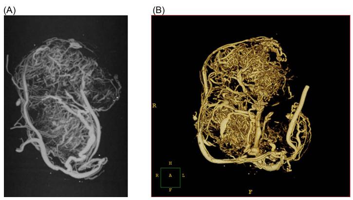
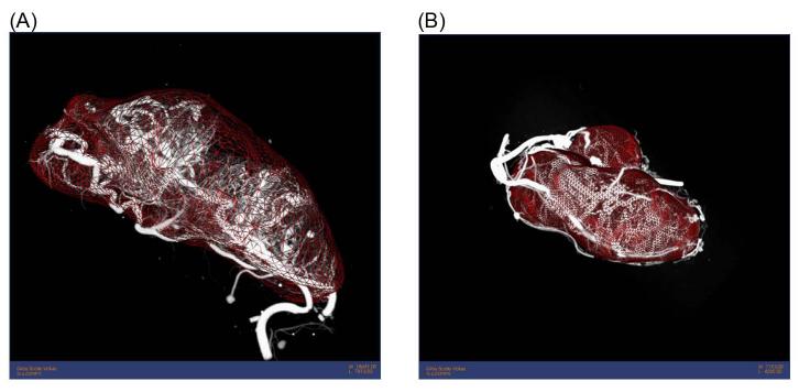
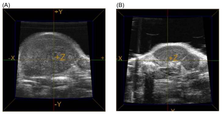
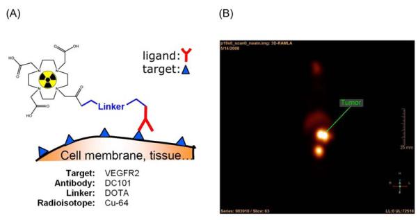
Vascular imaging of angiogenesis in mouse models of disease requires multi modal imaging hardware capable of targeting both structure and function at different physical scales. The three dimensional (3D) structure and function vascular information allows for accurate differentiation between biological processes. For example, image analysis of vessel development in angiogenesis vs. arteriogenesis enables more accurate detection of biological variation between subjects and more robust and reliable diagnosis of disease. In the recent years a number of micro imaging modalities have emerged in the field as preferred means for this purpose. They provide 3D volumetric data suitable for analysis, quantification, validation, and visualization of results in animal models. This review highlights the capabilities of microCT, ultrasound and microPET for multimodal imaging of angiogenesis and molecular vascular targets in a mouse model of tumor angiogenesis. The basic principles of the imaging modalities are described and experimental results are presented.
Figures
Similar articles
-
New trends in molecular imaging of tumor angiogenesis.Anticancer Agents Med Chem. 2008 Jun;8(5):497-522. doi: 10.2174/187152008784533026. Anticancer Agents Med Chem. 2008. PMID: 18537533 Review.
-
Imaging of tumor angiogenesis: current approaches and future prospects.Curr Pharm Des. 2006;12(21):2661-72. doi: 10.2174/138161206777698774. Curr Pharm Des. 2006. PMID: 16842165 Review.
-
Optical imaging and tumor angiogenesis.J Cell Biochem. 2003 Oct 15;90(3):484-91. doi: 10.1002/jcb.10630. J Cell Biochem. 2003. PMID: 14523982 Review.
-
[Applications of fluorescent molecular imaging in tumor detection].Sheng Wu Yi Xue Gong Cheng Xue Za Zhi. 2010 Oct;27(5):1152-7. Sheng Wu Yi Xue Gong Cheng Xue Za Zhi. 2010. PMID: 21089689 Review. Chinese.
-
Multimodal microvascular imaging reveals that selective inhibition of class I PI3K is sufficient to induce an antivascular response.Neoplasia. 2013 Jul;15(7):694-711. doi: 10.1593/neo.13470. Neoplasia. 2013. PMID: 23814482 Free PMC article.
Cited by
-
A new method for in vivo visualization of vessel remodeling using a near-infrared dye.Microcirculation. 2011 Apr;18(3):163-71. doi: 10.1111/j.1549-8719.2011.00085.x. Microcirculation. 2011. PMID: 21418375 Free PMC article.
-
Osmotic drug delivery to ischemic hindlimbs and perfusion of vasculature with microfil for micro-computed tomography imaging.J Vis Exp. 2013 Jun 29;(76):50364. doi: 10.3791/50364. J Vis Exp. 2013. PMID: 23852145 Free PMC article.
-
Evaluating the effect of Avastin on breast cancer angiogenesis using synchrotron radiation.Quant Imaging Med Surg. 2019 Mar;9(3):418-426. doi: 10.21037/qims.2019.03.09. Quant Imaging Med Surg. 2019. PMID: 31032189 Free PMC article.
-
Imaging stem cell therapy for the treatment of peripheral arterial disease.Curr Vasc Pharmacol. 2012 May;10(3):361-73. doi: 10.2174/157016112799959404. Curr Vasc Pharmacol. 2012. PMID: 22239638 Free PMC article. Review.
-
Micro computed tomography for vascular exploration.J Angiogenes Res. 2010 Mar 5;2:7. doi: 10.1186/2040-2384-2-7. J Angiogenes Res. 2010. PMID: 20298533 Free PMC article.
References
-
- Folkman J. How is blood vessel growth regulated in normal and neoplastic tissue? G.H.A. Clowes memorial Award lecture. Cancer Res. 1986 Feb;46(2):467–473. - PubMed
-
- Alon T, Hemo I, Itin A, Pe’er J, Stone J, Keshet E. Vascular endothelial growth factor acts as a survival factor for newly formed retinal vessels and has implications for retinopathy of prematurity. Nat Med. 1995 Oct;1(10):1024–1028. - PubMed
-
- Ferrara N. Role of vascular endothelial growth factor in regulation of physiological angiogenesis. Am J Physiol Cell Physiol. 2001 Jun;280(6):C1358–1366. - PubMed
-
- Ferrara N, Chen H, Davis-Smyth T, Gerber HP, Nguyen TN, Peers D, Chisholm V, Hillan KJ, Schwall RH. Vascular endothelial growth factor is essential for corpus luteum angiogenesis. Nat Med. 1998 Mar;4(3):336–340. - PubMed
-
- Folkman J. Angiogenesis in cancer, vascular, rheumatoid and other disease. Nat Med. 1995 Jan;1(1):27–31. - PubMed
Publication types
MeSH terms
Grants and funding
LinkOut - more resources
Full Text Sources
Medical
