Case Reports
. 2009 Jun;29(6):956-62.
doi: 10.1161/ATVBAHA.109.186577. Epub 2009 Mar 19.

Chylomicronemia with a mutant GPIHBP1 (Q115P) that cannot bind lipoprotein lipase

Affiliations
Case Reports

Chylomicronemia with a mutant GPIHBP1 (Q115P) that cannot bind lipoprotein lipase

Anne P Beigneux et al. Arterioscler Thromb Vasc Biol. 2009 Jun.

Abstract

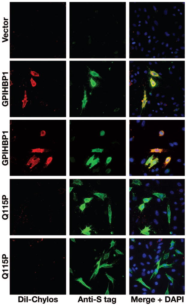
Objective: GPIHBP1 is an endothelial cell protein that binds lipoprotein lipase (LPL) and chylomicrons. Because GPIHBP1 deficiency causes chylomicronemia in mice, we sought to determine whether some cases of chylomicronemia in humans could be attributable to defective GPIHBP1 proteins.

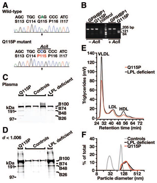
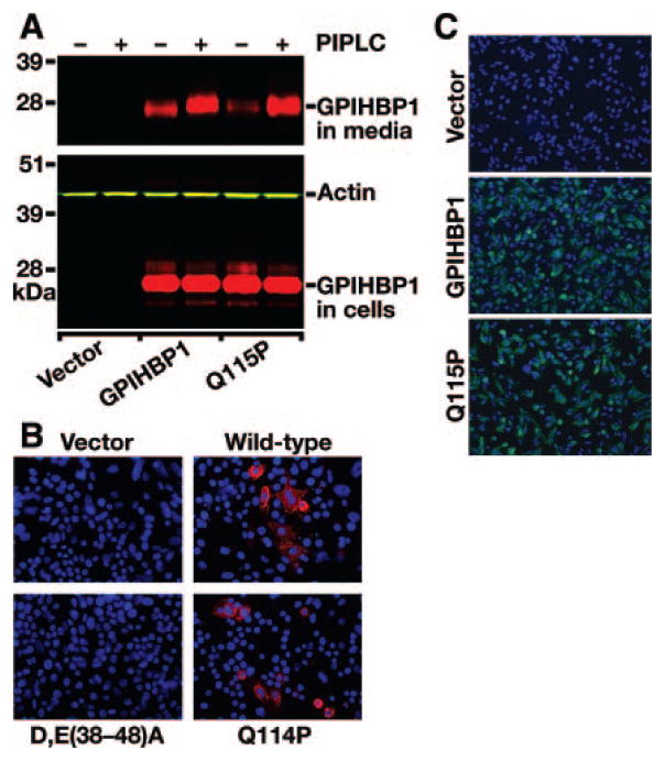
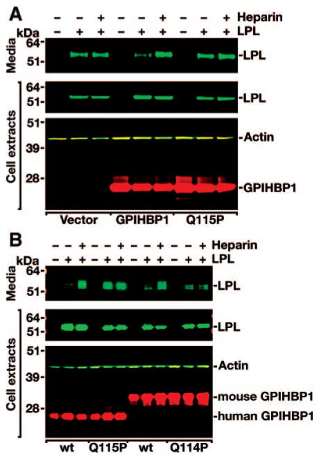
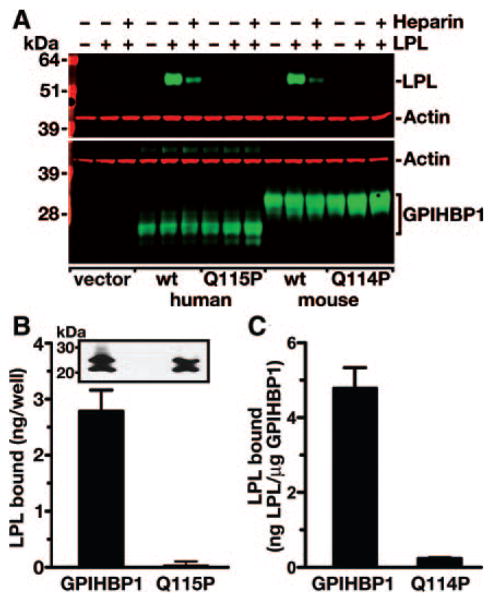
Methods and results: Patients with severe hypertriglyceridemia (n=60, with plasma triglycerides above the 95th percentile for age and gender) were screened for mutations in GPIHBP1. A homozygous GPIHBP1 mutation (c.344A>C) that changed a highly conserved glutamine at residue 115 to a proline (p.Q115P) was identified in a 33-year-old male with lifelong chylomicronemia. The patient had failure-to-thrive as a child but had no history of pancreatitis. He had no mutations in LPL, APOA5, or APOC2. The Q115P substitution did not affect the ability of GPIHBP1 to reach the cell surface. However, unlike wild-type GPIHBP1, GPIHBP1-Q115P lacked the ability to bind LPL or chylomicrons (d < 1.006 g/mL lipoproteins from Gpihbp1(-/-) mice). Mouse GPIHBP1 with the corresponding mutation (Q114P) also could not bind LPL.

Conclusions: A homozygous missense mutation in GPIHBP1 (Q115P) was identified in a patient with chylomicronemia. The mutation eliminated the ability of GPIHBP1 to bind LPL and chylomicrons, strongly suggesting that it caused the patient's chylomicronemia.


Figures

Figure 1
Figure 1
Identification of a homozygous mutation in GPIHBP1 (p.Q115P) in a young man with chylomicronemia. A, DNA sequence of GPIHBP1 exon 4 from a normolipidemic control subject (wild-type) and the Q115P homozygote (p.Q115P mutant). Nucleotide and amino acid sequences are shown above each chromatogram. The arrow indicates the nucleotide substitution (c.344A>C); this mutation creates a new AciI site. B, AciI digestion confirming the Q115P mutation. A 205-bp fragment of GPIHBP1 was amplified from genomic DNA with primers 5′-CCATCCTCAGCACTTGTTCCCCACTCCCC-3′ and 5′-CCTGCCCCCTTGCCTGTTGGG TCC-3′. AciI cleaves the DNA fragment from the Q115P homozygote. C and D, Increased amounts of apo-B48 in the plasma (C) and in the d<1.006 g/mL lipoproteins (D) of the Q115P proband, as judged by Western blots with an apo-B–specific monoclonal antibody (MB3). Samples from normolipidemic subjects and an LPL-deficient patient were included as controls. E, Distribution of triglycerides within the chylomicron-depleted lipoproteins of the proband (Q115P) and an LPL-deficient patient. Plasma lipoproteins were size-fractionated by fast protein liquid chromatography with a Superose 6 HR column. F, Distribution of lipoprotein sizes (as judged by dynamic laser light scattering) in the d<1.006 g/mL lipoproteins from the Q115P homozygote, an LPL-deficient patient, and 2 normolipidemic control subjects. 98.3% of the plasma lipoproteins in the proband had diameters of 94 to 265 nm.
Figure 2
Figure 2
Wild-type and mutant (Q115P) versions of GPIHBP1 reach the cell surface. A, Release of wild-type or mutant (Q115P) GPIHBP1 from the surface of cells after incubation of cells with a phosphatidylinositol-specific phospholipase C (PIPLC). CHO pgsA-745 cells were transiently transfected with S-protein–tagged GPIHBP1 constructs. The release of GPIHBP1 from the cell surface after PIPLC treatment (1 U/mL for 1 hour at 37°C) was assessed by Western blotting. B, Binding of antibodies against the acidic domain of mouse GPIHBP1 to GPIHBP1-transfected cells. CHO pgsA-745 cells were transfected with empty vector or vectors encoding wild-type mouse GPIHBP1, GPIHBP1-Q114P, or a mouse GPIHBP1 mutant [D,E(38-48)A] in which the aspartates and glutamates between residues 38 and 48 were changed to alanine. GPIHBP1 on the surface of nonpermeabilized cells was assessed by immunofluorescence microscopy with a rabbit antiserum against the acidic domain of mouse GPIHBP1 (red). The GPIHBP1-D,E(38-48)A mutant is not recognized by the antiserum. Cell nuclei were visualized with DAPI (blue). C, Binding of antibodies against the S-protein tag to cells expressing human GPIHBP1. CHO pgsA-745 cells were electroporated with empty vector or vectors encoding wild-type mutant (Q115P) GPIHBP1. GPIHBP1 on the surface of nonpermeabilized cells was assessed by immunofluorescence microscopy using a goat antiserum against the S-protein tag (green). Cell nuclei were visualized with DAPI (blue).
Figure 3
Figure 3
Decreased binding of LPL to human GPIHBP1-Q115P. A, Western blot representative of 7 independent experiments of cell extracts and cell culture medium from CHO pgsA-745 cells that had been transfected with a V5-tagged human LPL expression vector, alone or in combination with an empty vector, a vector for an S-protein–tagged wild-type human GPIHBP1, or a vector for a mutant human GPIHBP1 with the Q115P mutation. B, Western blot analysis of CHO pgsA-745 cells that had been transfected with a V5-tagged human LPL expression vector, alone or in combination with S-protein–tagged wild-type or mutant (Q115P) human GPIHBP1 constructs, or wild-type or mutant (Q114P) mouse GPIHBP1 constructs.
Figure 4
Figure 4
Decreased binding of exogenously added LPL to cells expressing wild-type or mutant versions of GPIHBP1. A, Binding of human LPL to CHO pgsA-745 cells that had been electroporated with S-protein–tagged wild-type or mutant GPIHBP1 (Q115P) expression vectors. 24 hour after the electroporation, V5-tagged human LPL was added to the cells in the presence or absence of heparin (500 U/mL). After washing the cells, the amount of LPL in cell extracts was assessed by Western blotting. B, Binding of avian LPL to CHO pgsA-745 cells that had been transiently transfected with wild-type or mutant (Q115P) human GPIHBP1 expression vectors. 24 hour after the transfection, cells were incubated for 2 hours with avian LPL (225 ng/well). After washing the cells, the amount of LPL bound to cells was quantified with an ELISA. Inset shows a Western blot demonstrating comparable levels of expression for wild-type GPIHBP1 and GPIHBP1-Q115P. C, Binding of avian LPL to CHO pgsA-745 cells that had been transiently transfected with wild-type or mutant (Q114P) mouse GPIHBP1 expression vectors. After incubating the cells for 2 hours with avian LPL (225 ng/well), the cells were washed, and the amount of LPL bound to cells was quantified with a sandwich ELISA. LPL binding was normalized to the amount of GPIHBP1 (as judged by an ELISA) in the same well (wild-type, 1.85±0.16 μg/well; Q114P, 1.87±0.47 μg/well). The studies shown in B and C were performed in 2 entirely independent experiments, and identical results were obtained.
Figure 5
Figure 5
Decreased binding of DiI-labeled chylomicrons to cells expressing GPIHBP1-Q115P. CHO pgsA-745 cells were transfected with wild-type or mutant (Q115P) S-protein–tagged human GPIHBP1 expression vectors. GPIHBP1 on the surface of cells was detected by immunofluorescence microscopy with an antibody against the S-protein tag (green). Chylomicron binding was detected with DiI-fluorescence (red). Cell nuclei were visualized with DAPI (blue).

Comment in

References

    1. Brunzell JD, Deeb SS. Familial lipoprotein lipase deficiency, apo C-II deficiency, and hepatic lipase deficiency. In: Scriver CR, Beaudet AL, Sly WS, Valle D, Childs B, Kinzler KW, Vogelstein B, editors. The Metabolic and Molecular Bases of Inherited Disease. New York: McGraw-Hill; 2001.
    1. Beigneux AP, Davies B, Gin P, Weinstein MM, Farber E, Qiao X, Peale P, Bunting S, Walzem RL, Wong JS, Blaner WS, Ding ZM, Melford K, Wongsiriroj N, Shu X, de Sauvage F, Ryan RO, Fong LG, Bensadoun A, Young SG. Glycosylphosphatidylinositol-anchored high density lipoprotein–binding protein 1 plays a critical role in the lipolytic processing of chylomicrons. Cell Metab. 2007;5:279–291. - PMC - PubMed
    1. Havel RJ, Kane JP. Introduction: Structure and metabolism of plasma lipoproteins. In: Scriver CR, Beaudet AL, Sly WS, Valle D, Childs B, Kinzler KW, Vogelstein B, editors. The Metabolic and Molecular Bases of Inherited Disease. New York: McGraw-Hill; 2001.
    1. Gin P, Yin L, Davies BSJ, Weinstein MM, Ryan RO, Bensadoun A, Fong LG, Young SG, Beigneux AP. The acidic domain of GPIHBP1 is important for the binding of lipoprotein lipase and chylomicrons. J Biol Chem. 2008;283:29554–29562. - PMC - PubMed
    1. Mallya M, Campbell RD, Aguado B. Characterization of the five novel Ly-6 superfamily members encoded in the MHC, and detection of cells expressing their potential ligands. Protein Sci. 2006;15:2244–2256. - PMC - PubMed

Publication types

MeSH terms