

Review
doi: 10.1161/ATVBAHA.108.165563.
Epub 2009 Mar 20.
Inflammation imaging in atherosclerosis
Affiliations
- PMID: 19304673
- PMCID: PMC2836586
- DOI: 10.1161/ATVBAHA.108.165563
Item in Clipboard
Review
Inflammation imaging in atherosclerosis
Arterioscler Thromb Vasc Biol.
2009 Jul.
Abstract
Inflammation is important at many stages of atherosclerotic plaque development. We highlight several imaging modalities that can quantify the degree of plaque inflammation noninvasively. Imaging of this type might allow testing of novel antiatherosclerosis drugs, identification of patients at risk of plaque rupture, and deeper insight into the biology of the disease. The imaging modalities are discussed in relation to their potential use in these areas.
Conflict of interest statement
Figures
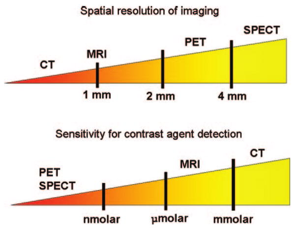
Illustration of the relative spatial resolution of common imaging techniques (top), along with their sensitivity values (bottom).
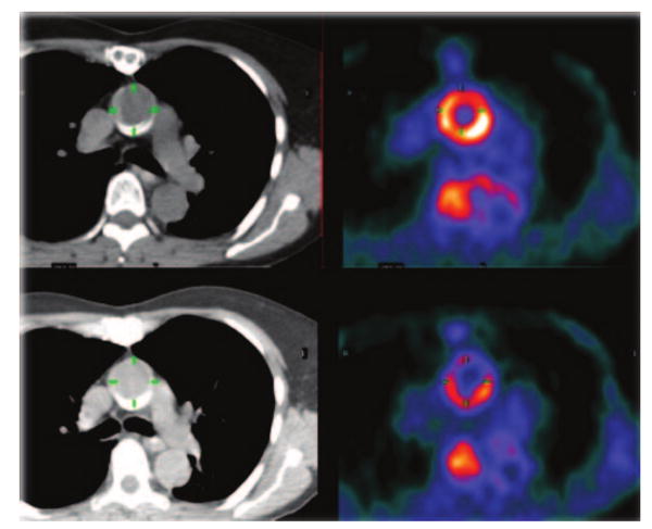
PET/CT image of aorta before (top) and during (bottom) antiatherosclerosis therapy. Note reduction in FDG uptake in the aortic wall.
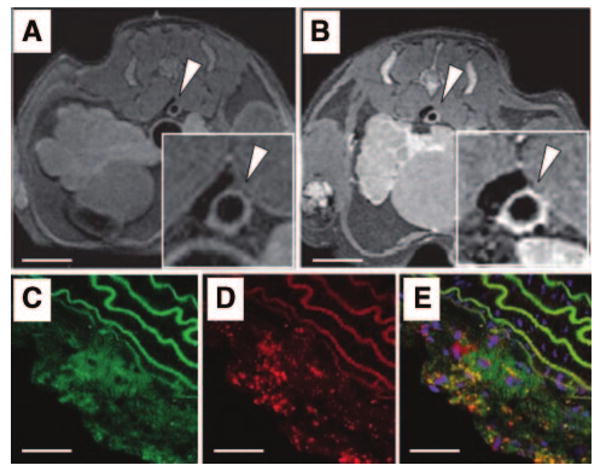
Detection of macrophages with gadolinium-containing micelles targeting macrophage scavenger receptors and MRI. MR axial views of an atherosclerotic plaque in the aorta of an atherosclerotic mice before (A) and 24 hours (B) after the intravenous injection of immuno-micelles targeting the macrophage scavenger receptors (Insets are magnification of the aorta; scale bar, 1 cm). A strong enhancement was detected with MRI in the aortic wall 24 hours after injection of immuno-micelles (B; white arrowhead). Confocal fluorescence microscopy of corresponding atherosclerotic plaque demonstrated colocalization between fluorescently labeled immunomicelles (C, green color) and anti-CD68 stained macrophages (D; red stain). Areas of yellow represent overlap of labeled immunomicelles and macrophage staining on the overlaid images (E). White scale bar, 250 μm. Adapted from Amirbekian et al.. Copyright © 2007 National Academy of Sciences, U.S.A.
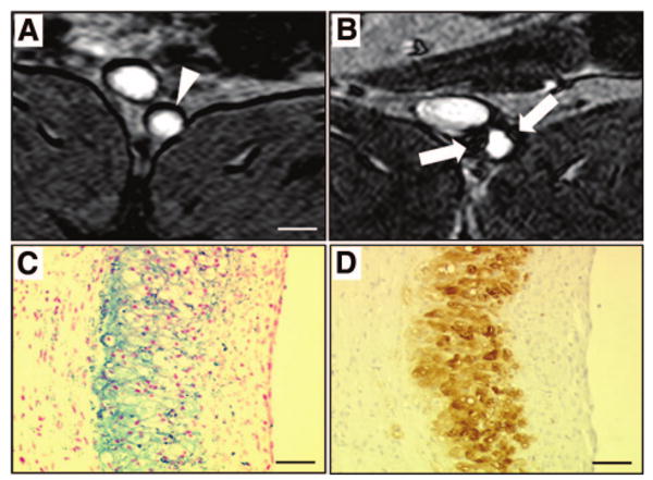
Detection of macrophages with USPIO-enhanced MRI. MR axial views of an atherosclerotic plaque in a rabbit aorta before (A) and 5 days (B) after the intravenous injection of ultrasmall superparamagnetic iron oxide nanoparticles (USPIO). Five days after the intravenous injection of USPIO, strong signal voids (white arrows) were detected in the aortic wall using T2*-weighted MR sequences. Note the dark artifacts that are not related to USPIO accumulation at the fat-water interfaces. On corresponding histological section, accumulation of iron oxide nanoparticles was identified in the atherosclerotic plaque with Perls stain (C; blue staining for iron) and colocalized with macrophage infiltration detected by immunohistotochemistry using a RAM-11 monoclonal antibody for rabbit macrophages (D; brown staining). White scale bar, 5 mm; black scale bar, 30 μm.
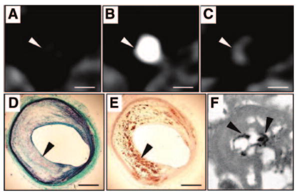
Detection of macrophages with N1177-enhanced CT. CT axial views of an atherosclerotic plaque in a rabbit aorta before (A), during (B), and 2 hours after (C) the intravenous injection of the contrast agent N1177. Note the strong enhancement of the aortic wall detected with CT 2 hours after the injection of N1177 (C; white arrowhead). On axial sections corresponding to the CT images, atherosclerotic plaque was characterized by a large lipid-rich core covered by a thin cap of collagen stained in green with Masson trichrome (D; black arrowhead) and intense macrophage infiltration in the lipid-rich core detected by immunohistochemistry for macrophages with a monoclonal RAM-11 antibody (E; black arrowhead). Numerous iodine particles (black arrowheads) were detected with transmission electron microscopy, next to lipid inclusions, in lysosomes of macrophages from atherosclerotic plaques (F). White scale bar, 5 mm; black scale bar, 1 mm. Adapted from Hyafil et al.
References
-
- Rosamond W, Flegal K, Friday G, Furie K, Go A, Greenlund K, Haase N, Ho M, Howard V, Kissela B, Kittner S, Lloyd-Jones D, McDermott M, Meigs J, Moy C, Nichol G, O'Donnell CJ, Roger V, Rumsfeld J, Sorlie P, Steinberger J, Thom T, Wasserthiel-Smoller S, Hong Y, for the American Heart Association Statistics Committee and Stroke Statistics Subcommittee Heart Disease and Stroke Statistics–2007 Update: A Report From the American Heart Association Statistics Committee and Stroke Statistics Subcommittee. Circulation. 2007;115:e69–e171. - PubMed
-
- Virmani R, Burke AP, Farb A, Kolodgie FD. Pathology of the vulnerable plaque. J Am Coll Cardiol. 2006;47:C13–C18. - PubMed
-
- Burke AP, Farb A, Malcom GT, Liang YH, Smialek J, Virmani R. Coronary risk factors and plaque morphology in men with coronary disease who died suddenly. N Engl J Med. 1997;336:1276–1282. - PubMed
-
- Crisby M, Nordin-Fredriksson G, Shah PK, Yano J, Zhu J, Nilsson J. Pravastatin treatment increases collagen content and decreases lipid content, inflammation, metalloproteinases, and cell death in human carotid plaques: implications for plaque stabilization. Circulation. 2001;103:926–933. - PubMed
-
- Naghavi M, Falk E, Hecht HS, Jamieson MJ, Kaul S, Berman DS, Fayad ZA, Budoff MJ, Rumberger J, Naqvi TZ, Shaw LJ, Faergeman O, Cohn J, Bahr R, Koenig W, Demirovic J, Arking D, Herrera VL, Badimon JJ, Goldstein JA, Rudy Y, Airaksinen J, Schwartz RS, Riley WA, Mendes RA, Douglas P, Shah PK, Force ST. From vulnerable plaque to vulnerable patient–Part III: Executive summary of the Screening for Heart Attack Prevention and Education (SHAPE) Task Force report. The Am J Cardiol. 2006;98:2H–15H. - PubMed
Publication types
MeSH terms
Substances
Grants and funding
LinkOut - more resources
Full Text Sources
Other Literature Sources
Medical
