. 2009 May;37(8):e62.
doi: 10.1093/nar/gkp176. Epub 2009 Mar 20.

Selection of aptamers for a protein target in cell lysate and their application to protein purification

Affiliations

Selection of aptamers for a protein target in cell lysate and their application to protein purification

Sahar Javaherian et al. Nucleic Acids Res. 2009 May.

Abstract

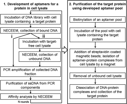
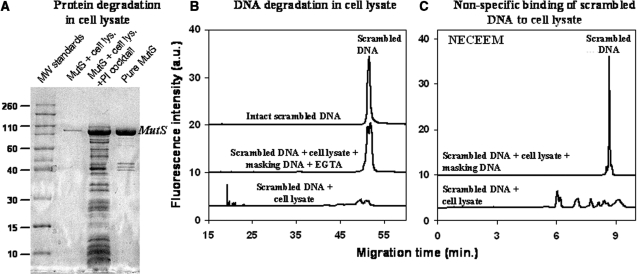
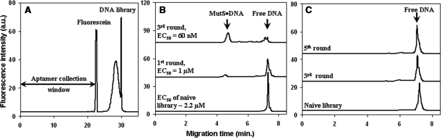
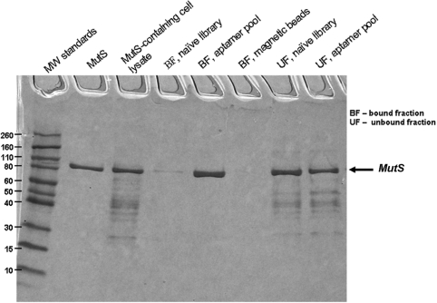
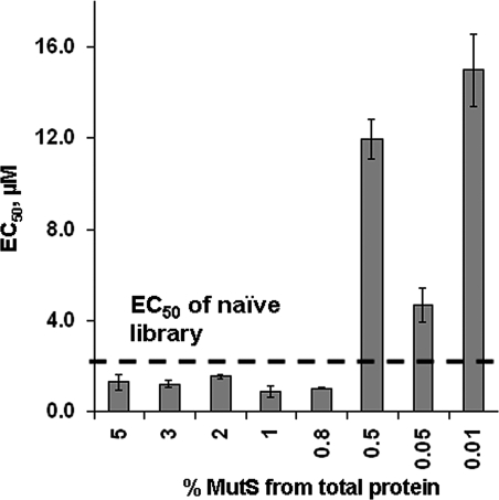
Functional genomics requires structural and functional studies of a large number of proteins. While the production of proteins through over-expression in cultured cells is a relatively routine procedure, the subsequent protein purification from the cell lysate often represents a significant challenge. The most direct way of protein purification from a cell lysate is affinity purification using an affinity probe to the target protein. It is extremely difficult to develop antibodies, classical affinity probes, for a protein in the cell lysate; their development requires a pure protein. Thus, isolating the protein from the cell lysate requires antibodies, while developing antibodies requires a pure protein. Here we resolve this loop problem. We introduce AptaPIC, Aptamer-facilitated Protein Isolation from Cells, a technology that integrates (i) the development of aptamers for a protein in cell lysate and (ii) the utilization of the developed aptamers for protein isolation from the cell lysate. Using MutS protein as a target, we demonstrate that this technology is applicable to the target protein being at an expression level as low as 0.8% of the total protein in the lysate. AptaPIC has the potential to considerably speed up the purification of proteins and, thus, accelerate their structural and functional studies.


Figures

Figure 1.
Figure 1.
Aptamer-facilitated protein isolation from cells (AptaPIC). (i) denotes aptamer selection by consecutive rounds of positive selection. (ii) denotes aptamer selection with alternating positive and negative selection.
Figure 2.
Figure 2.
Three processes that interfere with aptamer development for a target protein in cell lysate: protein degradation (A), DNA degradation (B) and binding of DNA by non-target components of cell lysate (C). (A) MutS degradation was analyzed by SDS–PAGE with Coomassie staining. The four lanes (from left to right) in the gel image correspond to: (i) molecular weight standards (×1000), (ii) MutS degraded by proteases in the cell lysate, (iii) MutS rescued from proteolysis in the cell lysate by a protease inhibitor (PI) cocktail, and (iv) pure MutS. The samples analyzed contained 972 µg/ml of cell lysate protein and 54 µg/ml of MutS. (B) DNA degradation was analyzed by gel capillary electrophoresis (Gel CE) using a scrambled 80-nt DNA as an experimental model. The three traces (from bottom to top) correspond to: (i) scrambled DNA degraded in cell lysate, (ii) scrambled DNA rescued from the degradation in the cell lysate by masking DNA and EGTA and (iii) scrambled DNA as a control. (C) Binding of DNA to non-target components of the cell lysate was studied by NECEEM using the scrambled DNA. The two traces (from bottom to top) in (C) correspond to: (i) scrambled DNA bound to non-target components of the cell lysate and/or partially degraded DNA and (ii) scrambled DNA rescued from binding and degradation by masking DNA. NECEEM was carried out in a 50-cm-long capillary at an electric field of 400 V/cm with 50 mM Tris–acetate at pH 8.3 as a run buffer.
Figure 3.
Figure 3.
Selection of aptamers for MutS in the E. coli cell lysate by consecutive rounds of positive selection. (A) Aptamer collection window for NECEEM-based selection of aptamers. The equilibrium mixture was injected into the capillary and electrophoresis was carried out in an 80-cm-long capillary at a 250 V/cm electric field with 50 mM Tris–acetate at pH 8.3 as a run buffer. The entire fraction migrating before fluorescein was collected for PCR amplification of DNA contained in it. (B) Progression of selection with regards to MutS. A naive DNA library and enriched aptamer pools were incubated with pure MutS and NECEEM was used to estimate EC50 values for 220 nM MutS. The three traces (from bottom to top) correspond to the mixture of MutS with (i) naive library, (ii) enriched DNA library after one round of aptamer selection and (iii) enriched DNA library after three rounds of aptamer selection. (C) Progression of selection with regards to the MutS-free cell lysate. The naive DNA library and enriched aptamer pools were incubated with MutS-free cell lysate and analyzed by NECEEM. The three traces (from bottom to top) correspond to mixtures of MutS-free cell lysate with: (i) naive library, (ii) enriched DNA library after three rounds of aptamer selection and (iii) enriched DNA library after five rounds of aptamer selection. In (B) and (C), NECEEM was carried out in a 50-cm-long capillary at an electric field of 400 V/cm with 50 mM Tris–acetate at pH 8.3 as a run buffer.
Figure 4.
Figure 4.
SDS–PAGE with Coomassie staining of affinity pull-down of MutS from the E. coli cell lysate using the aptamers developed by AptaPIC. The lanes from left to right correspond to: (i) molecular weight standards (×1000); (ii) pure MutS; (iii) MutS-containing cell lysate, the sample contained 91.25 µg/ml and 18 µg/ml of MutS; (iv) MutS purification using the naive DNA library, bound fraction; (v) MutS purification using the developed aptamer pool, bound fraction; (vi) fraction bound to streptavidin-coated magnetic beads; (vii) MutS purification using the naive DNA library, unbound fraction; and (viii) MutS purification using the developed aptamer pool, unbound fraction.
Figure 5.
Figure 5.
EC50 values for the interaction of MutS with enriched libraries obtained after one round of aptamer selection for cell lysate containing MutS at indicated percentage levels with respect to the total protein in the lysate. All EC50 values were measured for a MutS concentration of 220 nM. Errors are those of EC50 measurements and not the selection.

References

    1. Biswas I, Hsieh P. Identification and characterization of a thermostable MutS homolog from Thermus aquaticus. J. Biol. Chem. 1996;271:5040–5048. - PubMed
    1. Koles K, Irvine KD, Panin VM. Functional characterization of Drosophila sialyltransferase. J. Biol. Chem. 2004;279:4346–4357. - PubMed
    1. Arnau J, Lauritzen C, Petersen GE, Pedersen J. Current strategies for the use of affinity tags and tag removal for the purification of recombinant proteins. Protein Express. Purif. 2006;48:1–13. - PubMed
    1. Blecher SR, Howie R, Li S, Detmar J, Blahut LM. A new approach to immunological sexing of sperm. Theriogenology. 1999;52:1309–1321. - PubMed
    1. Ellington AD, Szostak JW. In vitro selection of RNA molecules that bind specific ligands. Nature. 1990;346:818–822. - PubMed

Publication types

MeSH terms