. 2009 Apr;136(8):1351-61.
doi: 10.1242/dev.027342.

Recruitment and maintenance of tendon progenitors by TGFbeta signaling are essential for tendon formation

Affiliations

Recruitment and maintenance of tendon progenitors by TGFbeta signaling are essential for tendon formation

Brian A Pryce et al. Development. 2009 Apr.

Abstract

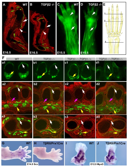
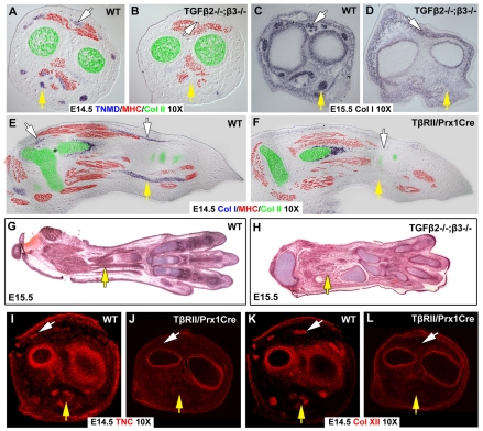
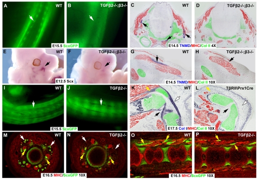
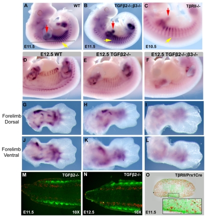
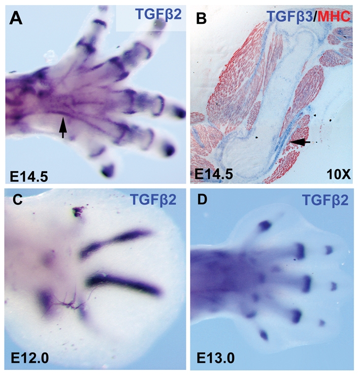
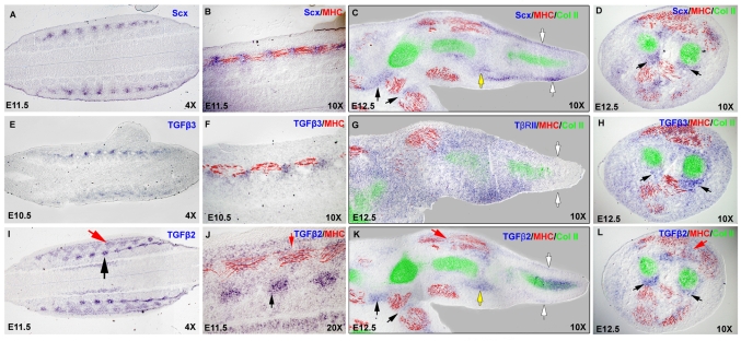
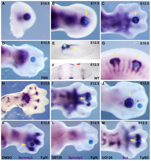
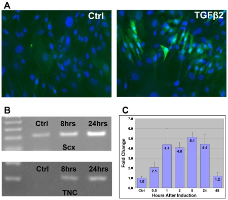
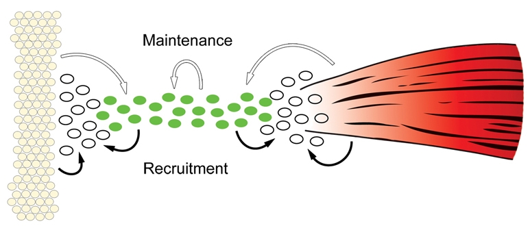
Tendons and ligaments mediate the attachment of muscle to bone and of bone to bone to provide connectivity and structural integrity in the musculoskeletal system. We show that TGFbeta signaling plays a major role in the formation of these tissues. TGFbeta signaling is a potent inducer of the tendon progenitor (TNP) marker scleraxis both in organ culture and in cultured cells, and disruption of TGFbeta signaling in Tgfb2(-/-);Tgfb3(-/-) double mutant embryos or through inactivation of the type II TGFbeta receptor (TGFBR2; also known as TbetaRII) results in the loss of most tendons and ligaments in the limbs, trunk, tail and head. The induction of scleraxis-expressing TNPs is not affected in mutant embryos and the tendon phenotype is first manifested at E12.5, a developmental stage in which TNPs are positioned between the differentiating muscles and cartilage, and in which Tgfb2 or Tgfb3 is expressed both in TNPs and in the differentiating muscles and cartilage. TGFbeta signaling is thus essential for maintenance of TNPs, and we propose that it also mediates the recruitment of new tendon cells by differentiating muscles and cartilage to establish the connections between tendon primordia and their respective musculoskeletal counterparts, leading to the formation of an interconnected and functionally integrated musculoskeletal system.


Figures

Fig. 1.
Fig. 1.
Loss of ScxGFP tendon signal in an allelic series of Tgfb2- and Tgfb3-null alleles. Tendons were visualized using the ScxGFP transgenic reporter and sections were counterstained with an MHC antibody. (A,B) Sagittal sections through the forelimbs of an E16.5 wild-type mouse embryo and a Tgfb2-/- littermate. White arrowheads indicate the normal and missing Deltoid tuberosity in A and B, respectively; white arrows indicate the Deltoid tendon and missing tendon in A and B, respectively. (C,D) The extensor tendons of an E15.5 wild-type embryo and a Tgfb2-/- littermate visualized using the ScxGFP tendon reporter. White arrows indicate the extensor digitorium tendon in C and missing extensors in D; white arrowheads indicate extensor tendons in the digits. (E) Schematic drawing of the forelimb. The positions of sections shown in F are marked, 1, digits; 2, metacarpals; 3, proximal to the wrist. (F) Transverse sections through the forelimb of E14.5 embryos from an allelic series of Tgfb2 and Tgfb3 mutant mice. The panels are sub-indexed with a letter to denote the genotype: a, wild type; b, Tgfb2-/-; c, Tgfb2-/-;Tgfb3-/+; d, Tgfb2-/-;Tgfb3-/-. The numeral in each panel denotes the corresponding plane of section in E. White arrows indicate extensor tendons; yellow arrows, flexor tendons; pink arrows, muscles. (G-J) Whole-mount ISH on forelimbs from wild-type and Tgfbr2Prx1Cre embryos with a Scx probe at E14.5 (G,H) and a Myod probe at E12.5 (I,J).
Fig. 2.
Fig. 2.
All limb tendons are lost in Tgfb2-/-;Tgfb3-/- and Tgfbr2Prx1Cre mutant embryos. Comparison of tendon markers in sections from the limbs of wild-type and TGFβ signaling mutants. In all panels white arrows indicate extensor tendons, yellow arrows indicate flexor tendons. (A-D) ISH with a Tnmd probe (A,B) and a collagen I probe (C,D) on transverse sections through the zeugopod of wild-type embryos and Tgfb2-/-;Tgfb3-/- littermates at E14.5 and E15.5. Sections in A and B were subsequently stained with antibodies for MHC (red) and collagen II (green). (E,F) ISH with a collagen I probe on sagittal sections through the limbs of an E14.5 wild-type embryo and a Tgfbr2Prx1Cre littermate was followed by antibody staining for MHC (red) and collagen II (green). (G,H) Hematoxylin and Eosin staining of planar sections through limbs of an E15.5 wild-type embryo and a Tgfb2-/-;Tgfb3-/- littermate. (I-L) Antibody staining for tenascin C (I,J) and collagen XII (K,L) on transverse sections through the zeugopod of a wild-type embryo (I,K) and a Tgfbr2Prx1Cre littermate (J,L).
Fig. 3.
Fig. 3.
Tendons and ligaments throughout the body are lost in TGFβ signaling mutants. (A,B) The ScxGFP tendon reporter in trunks of skinned E15.5 wild-type embryos and Tgfb2-/-;Tgfb3-/- littermates. Arrows indicate tendons. (C,D,G,H) Transverse sections through the heads of a wild-type embryo (C,G) and a Tgfb2-/-;Tgfb3-/- littermate (D,H) at E14.5 were processed for ISH for Tnmd, followed by immunostaining with antibodies to MHC (red) and collagen II (green). (C,D) Neck muscles. (G,H) The masseter muscle of the jaw. Arrows indicate actual and missing Tnmd signal. (E,F) Whole-mount ISH for Scx on heads of an E12.5 wild-type embryo and a Tgfb2-/-;Tgfb3-/- littermate. Arrows indicate Scx in the masseter. (I,J) ScxGFP in tails from an E15.5 wild-type embryo and a Tgfb2-/-;Tgfb3-/- littermate. Arrows indicate tendons. (K,L) Sagittal sections through the knees of E17.5 wild-type and Tgfbr2Prx1Cre embryos processed for Col1a1 ISH followed by immunostaining with antibodies to MHC (red) and collagen II (green). Yellow arrows, patellar tendons; white arrows, patellar ligament; black arrows, cruciate ligaments. (M-P) ScxGFP tendon reporter and antibodies to MHC (red) in transverse sections (M,N) and sagittal sections (O,P) through the tails of E16.5 wild-type (M,O) and Tgfb2-/- (N,P) embryos. White arrows, extrinsic tail tendons; yellow arrows, intrinsic muscles and tendons.
Fig. 4.
Fig. 4.
TNPs are lost in TGFβ signaling mutants at E12.5. (A-C) Whole-mount ISH for Scx on E11.5 wild-type (A), E11.5 Tgfb2-/-;Tgfb3-/- (B) and E10.5 Tgfbr2-/- (C) embryos. Yellow arrows, Scx expression in somites; red arrows, Scx expression in limb buds. The black line in A indicates the level of a frontal section through the trunk. (D-L) Whole-mount ISH for Scx on E12.5 whole embryos and dorsal and ventral forelimbs from wild-type (D,G,J), Tgfb2-/- (E,H,K) and Tgfb2-/-;Tgfb3-/- (F,I,L) embryos. (M,N) TUNEL staining (red) superimposed on ScxGFP signal (green) on frontal sections through the back (illustrated in A) of Tgfb2-/- embryos at E11.5 (M) and E12.5 (N). (O) BrdU staining superimposed on a ScxGFP signal in a transverse section through the limb bud of a E11.5 Tgfbr2Prx1Cre embryo. The inset is an enlargement of the boxed ventral Scx-positive domain.
Fig. 5.
Fig. 5.
Expression of Tgfb2 and Tgfb3. (A) Whole-mount ISH for Tgfb2 on an E14.5 hindlimb. (B) Section ISH for Tgfb3 followed by immunostaining with antibodies to MHC (red) on a sagittal section through the humerus of an E14.5 embryo. (C,D) Whole-mount ISH for Tgfb2 on forelimbs from embryos at E12.0 (C) and E13.0 (D).
Fig. 6.
Fig. 6.
Expression of Tgfb2 and Tgfb3 in and around the TNPs. (A-L) Section ISH for Scx (A-D), Tgfb3 (E,F,H), Tgfbr2 (G) and Tgfb2 (I-L) followed by immunostaining with antibodies to MHC (red) and collagen II (green). (A,B,E,F,I,J) Frontal sections through the back (illustrated in Fig. 4A) from embryos at E11.5 (A,B,I,J) and E10.5 (E,F). B, F and J are higher magnification images of A, E and I, respectively. Black arrows, early cartilage condensations; red arrows, myotome. (C,G,K) Sagittal sections thorough the forelimbs of an E12.5 embryo. White arrows, TNPs in the digits; red arrows, expression in muscle; black arrow, TNPs; yellow arrow, the wrist. (D,H,L) Transverse sections through the zeugopod of an E12.5 embryo at the level of the pronator quadratus muscle. Black arrows, TNPs; red arrow, expression in muscle.
Fig. 7.
Fig. 7.
Induction of Scx by TGFβ signaling in organ culture. (A-J) Whole mount ISH for Scx after 4-6 hours of incubation with Affigel beads saturated with 0.02 mg/ml TGFβ2 protein or PBS. Yellow arrows, induced Scx expression; red arrows, endogenous Scx expression. (A-C) TGFβ2 beads in forelimb buds from embryos at E10.5 (A), E11.5 (B) and E12.5 (C). (D) PBS control beads in a forelimb from an embryo at E12.5. (E) Transverse cryosection after Scx whole-mount ISH for an E12.5 limb incubated with a TGFβ2 bead. (F) Section ISH for Scx in a transverse section from an E12.5 wild-type forelimb in a position corresponding to the section in E. (G) TGFβ2 beads in the trunk of an E10.5 embryo. (H) Normal expression of Scx at E13.0. The red arrow highlights the sharp boundary for Scx expression at the metacarpal-phalangeal joint. (I) TGFβ2 beads at the level of the metacarpal-phalangeal joint at E12.5. (J) Distal and proximal TGFβ2 beads in an E12.5 limb. (K,L) Whole-mount ISH for Sprouty2 after a 6-hour incubation with an FGF4-loaded bead in the presence of 50 μM UO126 dissolved in DMSO (L), or just DMSO as control (K). (M) Whole-mount ISH for Scx after a 6-hour incubation with a TGFβ2 bead in the presence of 50 μM UO126.
Fig. 8.
Fig. 8.
Induction of early tendon markers by TGFβ signaling in tissue culture. (A) Mouse embryonic fibroblasts (MEFs) from ScxGFP embryos after a 24-hour incubation in culture medium alone (control) or supplemented with 0.3 nM TGFβ2 protein, counterstained with DAPI to detect the cell nuclei (blue). (B) Induction of Scx and tenascin C in C3H10T1/2 cells by TGFβ signaling. C3H10T1/2 cells were incubated in six-well plates with culture medium supplemented with 0.3 nM TGFβ2 protein for 8 hours or 24 hours. Semi-quantitative RT-PCR amplification (25 cycles) was performed to detect the relative levels of mRNA for Scx and tenascin C. (C) Changes in the levels of Scx transcript following a pulse of TGFβ activation. C3H10T1/2 cells incubated in six-well plates were supplemented with 0.3 nM TGFβ2 protein for one hour, after which the cells were washed and returned to regular medium. Cell were harvested at the indicated times after the initiation of the induction. Levels of Scx mRNA were determined by QRT-PCR and normalized to the levels of GAPDH, results of four separate experiments were averaged. Levels of Scx transcript are represented as fold change relative to non-induced cells.
Fig. 9.
Fig. 9.
TGFβ signaling promotes maintenance and recruitment of tendon progenitors. Tendon progenitors (green cells) that align between the cartilage condensations (yellow cells) and differentiating muscles (red) at E12.5 are dependent at this stage on TGFβ signaling (white arrows). This essential role for TGFβs from the muscles and cartilage (e.g. TGFβ2 which is expressed exclusively in these tissues in the somites) suggests also an affect on adjacent mesenchymal cells (white), possibly by recruiting them to the tendon cell fate (black arrows). Autocrine activity of TGFβs from the tendon progenitors plays a more minor role in progenitor maintenance. Although the loss of TGFβ3, which is expressed exclusively in tendon progenitors in the somites, does not result in tendon loss, the enhanced phenotype in double mutant Tgfb2-/-;Tgfb3-/- embryos shows that autocrine activity does contribute to progenitor maintenance (white arrow). It is also possible that TGFβs from the tendon progenitors contribute to the recruitment of neighboring mesenchymal cells (black arrows).

References

    1. Anderson, D. M., Arredondo, J., Hahn, K., Valente, G., Martin, J. F., Wilson-Rawls, J. and Rawls, A. (2006). Mohawk is a novel homeobox gene expressed in the developing mouse embryo. Dev. Dyn. 235, 792-801. - PubMed
    1. Baffi, M. O., Slattery, E., Sohn, P., Moses, H. L., Chytil, A. and Serra, R. (2004). Conditional deletion of the TGF-beta type II receptor in Col2a expressing cells results in defects in the axial skeleton without alterations in chondrocyte differentiation or embryonic development of long bones. Dev. Biol. 276, 124-142. - PubMed
    1. Baffi, M. O., Moran, M. A. and Serra, R. (2006). Tgfbr2 regulates the maintenance of boundaries in the axial skeleton. Dev. Biol. 296, 363-374. - PMC - PubMed
    1. Benjamin, M., Kaiser, E. and Milz, S. (2008). Structure-function relationships in tendons: a review. J. Anat. 212, 211-228. - PMC - PubMed
    1. Bi, Y., Ehirchiou, D., Kilts, T. M., Inkson, C. A., Embree, M. C., Sonoyama, W., Li, L., Leet, A. I., Seo, B. M., Zhang, L. et al. (2007). Identification of tendon stem/progenitor cells and the role of the extracellular matrix in their niche. Nat. Med. 13, 1219-1227. - PubMed

Publication types

MeSH terms