. 2009 May;10(5):480-7.
doi: 10.1038/ni.1720. Epub 2009 Mar 22.

Autophagy enhances the presentation of endogenous viral antigens on MHC class I molecules during HSV-1 infection

Affiliations

Autophagy enhances the presentation of endogenous viral antigens on MHC class I molecules during HSV-1 infection

Luc English et al. Nat Immunol. 2009 May.

Abstract

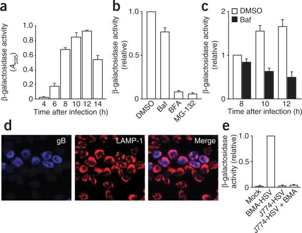
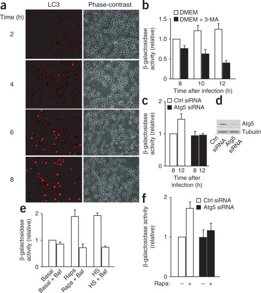
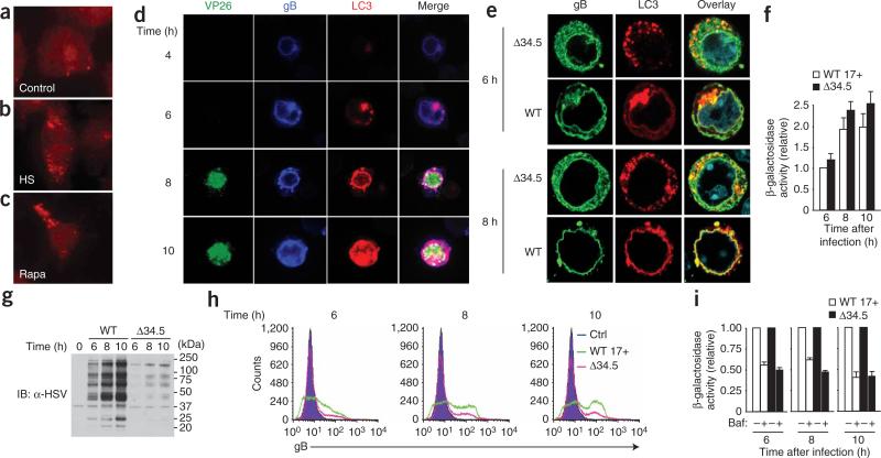
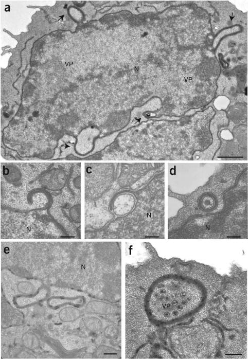
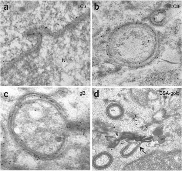
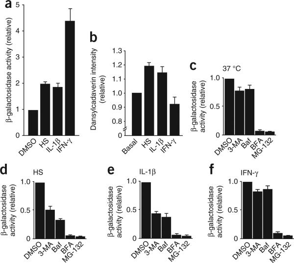
Viral proteins are usually processed by the 'classical' major histocompatibility complex (MHC) class I presentation pathway. Here we showed that although macrophages infected with herpes simplex virus type 1 (HSV-1) initially stimulated CD8(+) T cells by this pathway, a second pathway involving a vacuolar compartment was triggered later during infection. Morphological and functional analyses indicated that distinct forms of autophagy facilitated the presentation of HSV-1 antigens on MHC class I molecules. One form of autophagy involved a previously unknown type of autophagosome that originated from the nuclear envelope. Whereas interferon-gamma stimulated classical MHC class I presentation, fever-like hyperthermia and the pyrogenic cytokine interleukin 1beta activated autophagy and the vacuolar processing of viral peptides. Viral peptides in autophagosomes were further processed by the proteasome, which suggests a complex interaction between the vacuolar and MHC class I presentation pathways.


Figures

Figure 1
Figure 1
A vacuolar pathway participates in the processing of endogenous viral proteins for presentation on MHC class I molecules. (a) Activation of the gB-specific CD8+ T cell hybridoma (which expresses β-galactosidase as an indicator of T cell activation) by macrophages infected for various times (horizontal axis) with HSV-1, then incubated for 12 h at 37 °C with the hybridoma. A595, absorbance at 595 nm. (b) Activation of the hybridoma as described in a, with the addition of dimethyl sulfoxide (DMSO; negative control), bafilomycin A (Baf), brefeldin A (BFA) or MG-132 at 2 h after macrophage infection. (c) Activation of the hybridoma as described in a, with the addition of bafilomycin A at 2 h after macrophage infection. (d) Immunofluorescence microscopy of gB (blue) and LAMP-1 (red) in HSV-1-infected macrophages; pink indicates colocalization. Original magnification, ×40. (e) CD8+ T cell–stimulatory capacity (as described in a) of uninfected macrophages (Mock), of BMA (BMA-HSV) or J774 (J774-HSV) macrophages infected for 8 h with HSV-1, and of cocultures of J774 macrophages (H-2d) infected for 1 h with HSV-1, then mixed with uninfected BMA (H-2b) macrophages at a ratio of 1:1 and cultured together for 8 h (J774-HSV + BMA). Results in b,c,e are normalized to results obtained for CD8+ T cells stimulated with macrophages treated with DMSO (b,c) or infected BMA macrophages (e) and are presented in arbitrary units. Data are from one representative of three independent experiments (mean and s.d. of triplicate samples; a), are from three independent experiments (mean and s.e.m. of triplicate samples error bars; b,c,e) or are representative of three independent experiments (d).
Figure 2
Figure 2
Autophagy induced during HSV-1 infection contributes to the processing and presentation of endogenous viral antigens on MHC class I molecules. (a) Immunofluorescence microscopy of LC3 expression in macrophages infected for 2–8 h (left margin) with HSV-1. Original magnification, ×20. (b) Activation of the gB-specific CD8+ T cell hybridoma (described in Fig. 1a) by macrophages infected for various times (horizontal axis) with HSV-1, with (DMEM + 3-MA) or without (DMEM) the addition of 3-methyladenine 2 h after infection, then incubated for 12 h at 37 °C with the hybridoma. (c) Activation of the hybridoma (as described in b) by macrophages transfected for 60 h with control (Ctrl) siRNA or Atg5-specific siRNA, then infected for 8 h or 12 h with HSV-1. (d) Immunoblot analysis of Atg-5 in siRNA-treated macrophages. (e) Activation of the hybridoma (as described in b) by macrophages infected with HSV-1 and incubated at 37 °C (Basal), incubated for 12 h at 39 °C before being infected with HSV-1 (heat shock (HS)), or treated with rapamycin during HSV-1 infection (Rapa), with (+ Baf) or without the addition of bafilmycin A at 2 h after infection. (f) Activation of the hybridoma (as described in b) by macrophages transfected for 60 h with control siRNA or Atg5-specific siRNA, then infected for 8 h with HSV-1, with (+) or without (–) the addition of rapamycin at 2 h after infection. Results in b,c,e,f are normalized to results obtained for CD8+ T cells stimulated with macrophages infected for 8 h at 37 °C without further treatment (b,e) or infected macrophages treated with control siRNA (c,f) and are presented in arbitrary units. Data are representative of three independent experiments (a,d) or are from three independent experiments (mean and s.e.m. of triplicate samples; b,c,e,f).
Figure 3
Figure 3
Both gB and LC3 accumulate in perinuclear regions during HSV-1 infection. (a–c) Immunofluorescence microscopy of uninfected macrophages incubated at 37 °C (control; a), subjected to mild heat shock (b) or treated with rapamycin (c), then stained with anti-LC3. (d) Immunofluorescence microscopy of the expression of LC3, gB and GFP (VP26) by macrophages infected for various times (left margin) with HSV-1. White indicates colocalization. (e) Immunofluorescence microscopy of macrophages infected for 6 h or 8 h (left margin) with wild-type HSV-1 (WT) or HSV-1 lacking ICP34.5 (Δ34.5). Blue, staining of nuclei with DAPI (4,6-diamidino-2-phenylindole). Original magnification, ×100 (a–c) or ×63 (d,e). Results in a–e are representative of three independent experiments. (f) Activation of the gB-specific CD8+ T cell hybridoma (as described in Fig. 1a) by macrophages infected for various times (horizontal axis) with wild-type HSV-1 strain 17+ (WT 17+) or Δ34.5 HSV-1. Data are from three independent experiments (mean and s.e.m. of triplicate samples). (g,h) Immunoblot analysis (IB; g) and flow cytometry (h) of the expression of HSV-1 proteins (g) and gB (h) in macrophages infected for various times (above lanes (g) or plots (h)) with wild-type or Δ34.5 HSV-1. Ctrl, control (uninfected BMA macrophages; h). Data are representative of two (g) or three (h) independent experiments. (i) Activation of the gB-specific CD8+ T cell hybridoma (as described in Fig. 1a) by macrophages infected for various times (below graph) with wild-type or Δ34.5 HSV-1, with (+) or without (–) the addition of bafilomycin A at 2 h after infection. Results in f,i are normalized to results obtained for CD8+ T cells stimulated with macrophages infected for 6 h with wild-type virus (f) or with infected macrophages incubated without bafilomycin (i) and are presented in arbitrary units. Data are from three independent experiments (mean and s.e.m. of triplicate samples).
Figure 4
Figure 4
HSV-1 induces the formation of autophagosome-like structures from the nuclear envelopes of infected macrophages. Electron microscopy of macrophages 10 h after infection with HSV-1. (a) Arrows indicate membrane-coiled structures emerging from the nucleus of an infected cell. (b–d) Four-layered membrane structures formed by coiling of the nuclear membrane. (e,f) Glucose-6-phosphatase (black deposits) on autophagosome-like structures emerging from the nuclear envelope or free in the cytoplasm, and viral capsids in the cytoplasm engulfed in the lumen of an autophagosome-like compartment. N, nucleus; VP, viral particles. Scale bars, 1 μm (a), 0.25 μm (b–d,f) or 0.4 μm (e). Images are representative of three independent experiments with at least 100 cell profiles in each.
Figure 5
Figure 5
The four-layered membrane structures that emerge from the nuclear envelope have autophagosome-like features. Immunoelectron microscopy of macrophages 10 h after infection with HSV-1. (a–c) Accumulation of LC3 (a,b) and gB (c). (d) Fusion of four-layered membrane structures and lysosomes preloaded with bovine serum albumin–gold (BSA-gold; black dots). Original magnification, ×54,800 (a,b), ×69,000 (c) or ×38,000 (d). Images are representative of three (a–c) or two (d) independent experiments.
Figure 6
Figure 6
Involvement of lytic vacuolar compartments in the processing and presentation of endogenous antigens on MHC class I molecules after treatment with proinflammatory cytokines. (a) Activation of the gB-specific CD8+ T cell hybridoma (as described in Fig. 1a) by macrophages exposed to DMSO (negative control), mild heat shock, IL-1β or IFN-γ. (b) Dansylcadaverin staining of untreated macrophages (Basal) or macrophages exposed to mild heat shock, IFN-γ or IL-1β and infected for 8 h with wild-type HSV-1, normalized to the signal obtained in basal conditions and presented in arbitrary units. (c–f) Activation of the gB-specific CD8+ T cell hybridoma (as described in Fig. 1a) by macrophages incubated at 37 °C (c) or exposed to mild heat shock (d), IL-1β (e) or IFN-γ (f) and infected for 8 h with wild-type HSV-1 with the addition of DMSO (negative control), 3-methyladenine, bafilomycin, brefeldin A or MG-132 at 2 h after infection. Results in a,c–f are normalized to results obtained for CD8+ T cells stimulated with macrophages incubated with DMSO in each condition and are presented in arbitrary units. Data are from three independent experiments (mean and s.e.m. of triplicate samples).

References

    1. Bevan MJ. Cross-priming for a secondary cytotoxic response to minor H antigens with H-2 congenic cells which do not cross-react in the cytotoxic assay. J. Exp. Med. 1976;143:1283–1288. - PMC - PubMed
    1. Deretic V. Autophagy in innate and adaptive immunity. Trends Immunol. 2005;26:523–528. - PubMed
    1. Levine B, Deretic V. Unveiling the roles of autophagy in innate and adaptive immunity. Nat. Rev. Immunol. 2007;7:767–777. - PMC - PubMed
    1. Schmid D, Munz C. Innate and adaptive immunity through autophagy. Immunity. 2007;27:11–21. - PMC - PubMed
    1. Andrade RM, Wessendarp M, Gubbels MJ, Striepen B, Subauste CS. CD40 induces macrophage anti-Toxoplasma gondii activity by triggering autophagy-dependent fusion of pathogen-containing vacuoles and lysosomes. J. Clin. Invest. 2006;116:2366–2377. - PMC - PubMed

Publication types

MeSH terms