

Molecular docking and ligand specificity in fragment-based inhibitor discovery
- PMID: 19305397
- PMCID: PMC4006998
- DOI: 10.1038/nchembio.155
Molecular docking and ligand specificity in fragment-based inhibitor discovery
Abstract
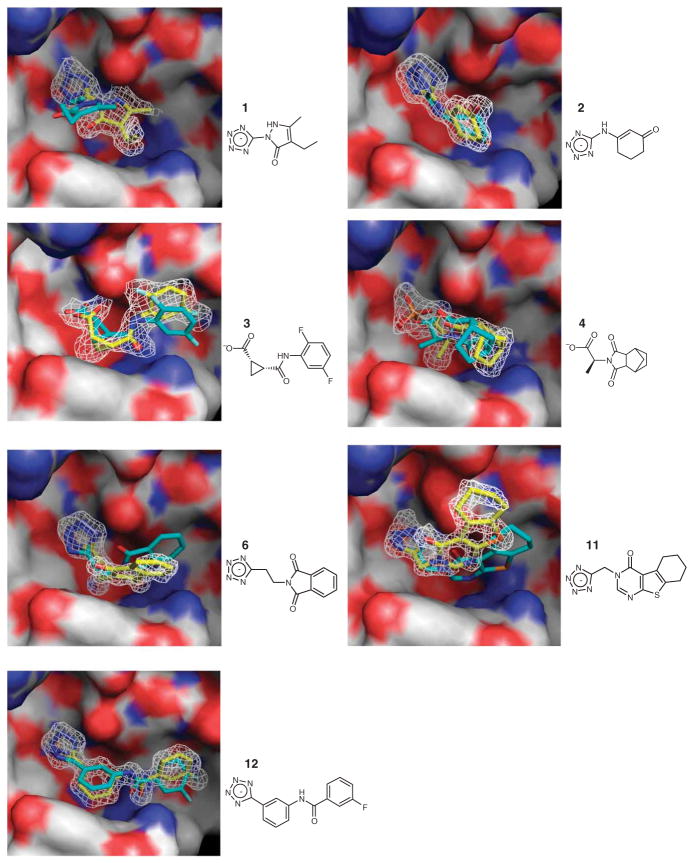
Fragment screens have successfully identified new scaffolds in drug discovery, often with relatively high hit rates (5%) using small screening libraries (1,000-10,000 compounds). This raises two questions: would other noteworthy chemotypes be found were one to screen all commercially available fragments (>300,000), and does the success rate imply low specificity of fragments? We used molecular docking to screen large libraries of fragments against CTX-M beta-lactamase. We identified ten millimolar-range inhibitors from the 69 compounds tested. The docking poses corresponded closely to the crystallographic structures subsequently determined. Notably, these initial low-affinity hits showed little specificity between CTX-M and an unrelated beta-lactamase, AmpC, which is unusual among beta-lactamase inhibitors. This is consistent with the idea that the high hit rates among fragments correlate to a low initial specificity. As the inhibitors were progressed, both specificity and affinity rose together, yielding to our knowledge the first micromolar-range noncovalent inhibitors against a class A beta-lactamase.
Figures

Comment in
-
Fragment-based drug discovery takes a virtual turn.Nat Chem Biol. 2009 May;5(5):274-5. doi: 10.1038/nchembio0509-274. Nat Chem Biol. 2009. PMID: 19377449 No abstract available.
References
-
- Rees DC, Congreve M, Murray CW, Carr R. Fragment-based lead discovery. Nat Rev Drug Discov. 2004;3:660–672. - PubMed
-
- Congreve M, Chessari G, Tisi D, Woodhead AJ. Recent developments in fragment-based drug discovery. J Med Chem. 2008;51:3661–3680. - PubMed
-
- Murray CW, et al. Application of fragment screening by X-ray crystallography to beta-secretase. J Med Chem. 2007;50:1116–1123. - PubMed
-
- Haydon DJ, et al. An inhibitor of FtsZ with potent and selective anti-staphylococcal activity. Science. 2008;321:1673–1675. - PubMed
-
- Card GL, et al. A family of phosphodiesterase inhibitors discovered by cocrystallography and scaffold-based drug design. Nat Biotechnol. 2005;23:201–207. - PubMed
Publication types
MeSH terms
Substances
Associated data
- Actions
- Actions
- Actions
- Actions
- Actions
- Actions
- Actions
- PubChem-Substance/57287690
- PubChem-Substance/57287691
- PubChem-Substance/57287692
- PubChem-Substance/57287693
- PubChem-Substance/57287694
- PubChem-Substance/57287695
- PubChem-Substance/57287696
- PubChem-Substance/57287697
- PubChem-Substance/57287698
- PubChem-Substance/57287699
- PubChem-Substance/57287700
- PubChem-Substance/57287701
- PubChem-Substance/57287702
- PubChem-Substance/57287703
- PubChem-Substance/57287704
- PubChem-Substance/57287705
- PubChem-Substance/57287706
- PubChem-Substance/57287707
- PubChem-Substance/57287708
- PubChem-Substance/57287709
- PubChem-Substance/57287710
- PubChem-Substance/57287711
- PubChem-Substance/57287712
- PubChem-Substance/57287713
- PubChem-Substance/57287714
- PubChem-Substance/57287715
- PubChem-Substance/57287716
- PubChem-Substance/57287717
- PubChem-Substance/57287718
- PubChem-Substance/57287719
- PubChem-Substance/57287720
- PubChem-Substance/57287721
- PubChem-Substance/57287722
- PubChem-Substance/57287723
- PubChem-Substance/57287724
- PubChem-Substance/57287725
- PubChem-Substance/57287726
- PubChem-Substance/57287727
- PubChem-Substance/57287728
- PubChem-Substance/57287729
- PubChem-Substance/57287730
- PubChem-Substance/57287731
- PubChem-Substance/57287732
- PubChem-Substance/57287733
- PubChem-Substance/57287734
- PubChem-Substance/57287735
- PubChem-Substance/57287736
- PubChem-Substance/57287737
- PubChem-Substance/57287738
- PubChem-Substance/57287739
- PubChem-Substance/57287740
- PubChem-Substance/57287741
- PubChem-Substance/57287742
- PubChem-Substance/57287743
- PubChem-Substance/57287744
- PubChem-Substance/57287745
- PubChem-Substance/57287746
- PubChem-Substance/57287747
- PubChem-Substance/57287748
- PubChem-Substance/57287749
- PubChem-Substance/57287750
- PubChem-Substance/57287751
- PubChem-Substance/57287752
- PubChem-Substance/57287753
- PubChem-Substance/57287754
- PubChem-Substance/57287755
- PubChem-Substance/57287756
- PubChem-Substance/57287757
- PubChem-Substance/57287758
- PubChem-Substance/57287759
- PubChem-Substance/57287760
- PubChem-Substance/57287761
- PubChem-Substance/57287762
- PubChem-Substance/57287763
- PubChem-Substance/57287764
- PubChem-Substance/57287765
- PubChem-Substance/57287766
- PubChem-Substance/57287767
- PubChem-Substance/57287768
- PubChem-Substance/57287769
- PubChem-Substance/57287770
- PubChem-Substance/57287771
- PubChem-Substance/57287772
- PubChem-Substance/57287773
- PubChem-Substance/57287774
- PubChem-Substance/57287775
- PubChem-Substance/57287776
- PubChem-Substance/57287777
- PubChem-Substance/57287778
- PubChem-Substance/57287779
- PubChem-Substance/57287780
- PubChem-Substance/57287781
- PubChem-Substance/57287782
- PubChem-Substance/57287783
- PubChem-Substance/57287784
- PubChem-Substance/57287785
- PubChem-Substance/57287786
- PubChem-Substance/57287787
- PubChem-Substance/57287788
- PubChem-Substance/57287789
- PubChem-Substance/57287790
- PubChem-Substance/57287791
- PubChem-Substance/57287792
- PubChem-Substance/57287793
- PubChem-Substance/57287794
- PubChem-Substance/57287795
- PubChem-Substance/57287796
- PubChem-Substance/57287797
- PubChem-Substance/57287798
- PubChem-Substance/57287799
- PubChem-Substance/57287800
- PubChem-Substance/57287801
- PubChem-Substance/57287802
- PubChem-Substance/57287803
- PubChem-Substance/57287804
Grants and funding
LinkOut - more resources
Full Text Sources
Other Literature Sources
Chemical Information
Research Materials
