. 2009;4(3):e4954.
doi: 10.1371/journal.pone.0004954. Epub 2009 Mar 23.

Glucose amplifies fatty acid-induced endoplasmic reticulum stress in pancreatic beta-cells via activation of mTORC1

Affiliations

Glucose amplifies fatty acid-induced endoplasmic reticulum stress in pancreatic beta-cells via activation of mTORC1

Etti Bachar et al. PLoS One. 2009.

Abstract

Background: Palmitate is a potent inducer of endoplasmic reticulum (ER) stress in beta-cells. In type 2 diabetes, glucose amplifies fatty-acid toxicity for pancreatic beta-cells, leading to beta-cell dysfunction and death. Why glucose exacerbates beta-cell lipotoxicity is largely unknown. Glucose stimulates mTORC1, an important nutrient sensor involved in the regulation of cellular stress. Our study tested the hypothesis that glucose augments lipotoxicity by stimulating mTORC1 leading to increased beta-cell ER stress.

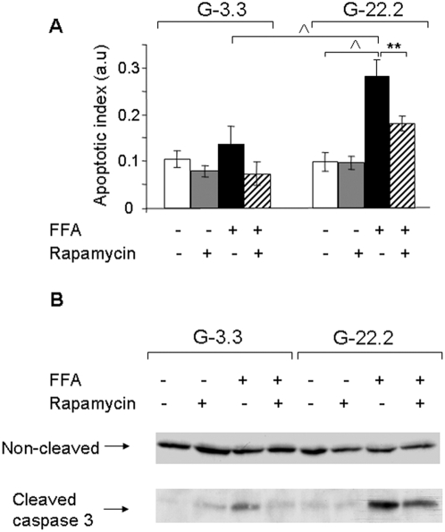
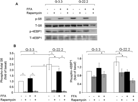
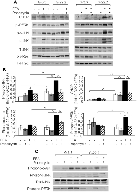
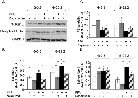
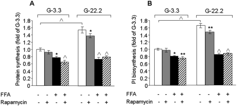
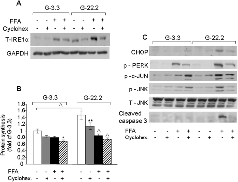
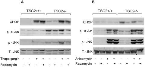
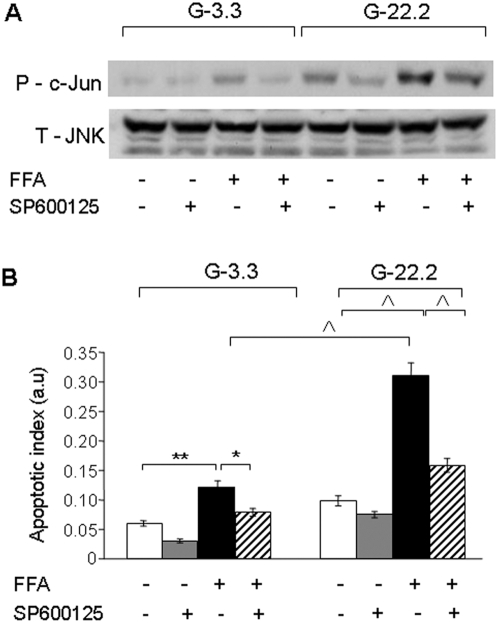
Principal findings: We found that glucose amplifies palmitate-induced ER stress by increasing IRE1alpha protein levels and activating the JNK pathway, leading to increased beta-cell apoptosis. Moreover, glucose increased mTORC1 activity and its inhibition by rapamycin decreased beta-cell apoptosis under conditions of glucolipotoxicity. Inhibition of mTORC1 by rapamycin did not affect proinsulin and total protein synthesis in beta-cells incubated at high glucose with palmitate. However, it decreased IRE1alpha expression and signaling and inhibited JNK pathway activation. In TSC2-deficient mouse embryonic fibroblasts, in which mTORC1 is constitutively active, mTORC1 regulated the stimulation of JNK by ER stressors, but not in response to anisomycin, which activates JNK independent of ER stress. Finally, we found that JNK inhibition decreased beta-cell apoptosis under conditions of glucolipotoxicity.

Conclusions/significance: Collectively, our findings suggest that mTORC1 mediates glucose amplification of lipotoxicity, acting through activation of ER stress and JNK. Thus, mTORC1 is an important transducer of ER stress in beta-cell glucolipotoxicity. Moreover, in stressed beta-cells mTORC1 inhibition decreases IRE1alpha protein expression and JNK activity without affecting ER protein load, suggesting that mTORC1 regulates the beta-cell stress response to glucose and fatty acids by modulating the synthesis and activity of specific proteins involved in the execution of the ER stress response. This novel paradigm may have important implications for understanding beta-cell failure in type 2 diabetes.


Conflict of interest statement

Competing Interests: The Israel Science Foundation, an Israeli National non-profit organization and a major source of competitive grant funding, supported part of the cost involved in performing the reseach. The Foundation was not involved in the decision to submit the work for publication.

Figures

Figure 1
Figure 1. Effect of mTORC1 inhibition by rapamycin on glucose and palmitate-induced β-cell apoptosis.
INS-1E cells were incubated at 3.3 and 22.2 mmol/l glucose with 0.5% BSA with and without 0.5 mmol/l palmitate and 50 nmol/l rapamycin for 16 h. Apoptosis was assessed using the Cell Death ELISAPLUS assay (Roche Diagnostics) (A) and by Western blot for cleaved caspase 3 (B). Results are expressed as means±SE of 4 individual experiments, each performed in triplicates (A). A representative gel of 3 individual experiments showing the expression of cleaved and uncleaved caspase 3 is presented (B). ** p<0.01, p<0.001 for the difference between the indicated groups.
Figure 2
Figure 2. Effects of glucose, palmitate and rapamycin on mTORC1 signaling in β-cells.
INS-1E cells were incubated overnight in RPMI medium containing 3.3 mmol/l glucose and 0.5% BSA without serum and then at 3.3 and 22.2 mmol/l glucose with and without 0.5 mmol palmitate and 50 nmol/l rapamycin for 4 h. S6(Ser235/236) and 4EBP1 phosphorylation were analyzed by Western blot. (A) A representative gel showing total and phosphorylated S6 and 4EBP1 is presented. (B) Quantification of S6 and 4EBP1 phosphorylation. Results are expressed as means±SE of 6 individual experiments. * p<0.05, p<0.001 for the difference between the indicated groups.
Figure 3
Figure 3. Effect of mTORC1 inhibition by rapamycin on glucose and palmitate-induced ER stress in β-cells and islets.
INS-1E cells were incubated at 3.3 and 22.2 mmol/l glucose with and without 0.5 mmol/l palmitate and 50 nmol/l rapamycin for 4 and 16 h; P. obesus islets were similarly treated for 24 h. Control incubations contained 0.5% BSA. ER stress was assessed by Western blot analysis for different ER stress markers. (A) A representative gel showing CHOP, phospho-PERK, phospho-c-Jun, phospho- and total JNK, phospho- and total eIF2α in INS-1E cells is presented. (B) Quantification of c-Jun, JNK, and PERK phosphorylation and CHOP expression at 16 h. Results are expressed as means±SE of 4 individual experiments. (C) A representative gel of 3 individual experiments showing phospho-c-Jun, phospho-PERK and phospho- and total JNK in P. obesus islets. * p<0.05, p<0.001 for the difference between the indicated groups or between the indicated groups and untreated controls at the same glucose concentration.
Figure 4
Figure 4. Effects of glucose, palmitate and rapamycin on IRE1α expression and activity.
INS-1E cells were incubated at 3.3 and 22.2 mmol/l glucose with and without 0.5 mmol/l palmitate and 50 nmol/l rapamycin for 16 h. (A) A representative gel showing total and phospho-IRE1α. (B, C) Quantification of IRE1α protein and mRNA levels, respectively. (D) Spliced Xbp-1 levels assessed by quantitative real-time PCR. Results are expressed as means±SE of 4–5 individual experiments. * p<0.05, p<0.001 for the difference between the indicated groups or between the indicated groups and untreated controls at the same glucose concentration.
Figure 5
Figure 5. Effects of glucose, palmitate and rapamycin on total protein and proinsulin biosynthesis.
INS-1E cells were incubated for 16 h at 3.3 and 22.2 mmol/l glucose with 0.5% BSA with and without 0.5 mmol/l palmitate and 50 nmol/l rapamycin. The last 2 h of the incubations were performed in KRBH-BSA buffer containing similar treatments and 10 µCi L-[2, 3, 4, 5-3H]leucine. After a 2-h incubation at 37°C, leucine incorporation was terminated by ice-cold wash-out in glucose-free KRBH-BSA buffer. Total protein synthesis (A) was determined by trichloroacetic acid precipitation. Proinsulin (PI) biosynthesis (B) was determined by immunoprecipitation with anti-insulin serum. Results are expressed as means±SE of 3 individual experiments, each performed in triplicates. * p<0.05, ** p<0.01, p<0.001 for the difference between the indicated groups or between the indicated groups and untreated controls at the same glucose concentration.
Figure 6
Figure 6. Effects of cycloheximide on IRE1α protein levels, total protein synthesis and different markers of ER stress in INS-1E cells treated with palmitate.
INS-1E cells were incubated at 3.3 and 22.2 mmol/l glucose with and without 0.5 mmol/l palmitate and 20 nmol/l cycloheximide for 16 h. (A) IRE1α expression was analyzed by Western blot. A representative gel of 4 individual experiments is shown. (B) Total protein synthesis was determined as described in Figure 5. Results are expressed as means±SE of 3 individual experiments, each performed in triplicates. (C) Western blot analysis for different ER stress and apoptosis markers. A representative gel of 4 individual experiments showing CHOP, phospho-PERK, phospho-c-Jun, phospho- and total JNK and cleaved caspase 3 is presented. * p<0.05, ** p<0.01, p<0.001 for the difference between the indicated groups or between the indicated groups and untreated controls at the same glucose concentration.
Figure 7
Figure 7. Effects of mTORC1 on ER stress-dependent and -independent JNK activation.
TSC2-deficient and wild-type mouse embryonic fibroblasts were treated with 300 nmol/l thapsigargin for 24 h (A) or with 200 nmol/l anisomycin for 30 min (B) with and without 50 nmol/l rapamycin, as described under Materials and Methods. JNK and c-Jun phosphorylation were analyzed by Western blot. Representative gels of 3 individual experiments showing CHOP, phoshpo-c-Jun, total and phospho-JNK are presented.
Figure 8
Figure 8. Effect of JNK inhibition on glucose and palmitate-induced β-cell apoptosis.
INS-1E cells were incubated for 16 h at 3.3 and 22.2 mmol/l glucose with 0.5% BSA with and without 0.5 mmol/l palmitate and 20 nmol/l of the JNK inhibitor SP600125. (A) Inhibition of JNK activation was studied by Western blot for phospho-c-Jun normalized to total JNK. A representative gel of 3 separate experiments is shown. The effects of JNK inhibition on apoptosis was analyzed using the apoptosis ELISA assay (B). Quantification of 3 individual experiments, each performed in triplicates is shown. * p<0.05, ** p<0.01, p<0.001 for the difference between the indicated groups.

References

    1. Poitout V, Robertson RP. Glucolipotoxicity: Fuel excess and beta-cell dysfunction. Endocr Rev. 2008;29:351–366. - PMC - PubMed
    1. Goh TT, Mason TM, Gupta N, So A, Lam TK, et al. Lipid-induced beta-cell dysfunction in vivo in models of progressive beta-cell failure. Am J Physiol Endocrinol Metab. 2007;292:E549–E560. - PubMed
    1. Maedler K, Spinas GA, Dyntar D, Moritz W, Kaiser N, et al. Distinct effects of saturated and monounsaturated fatty acids on beta-cell turnover and function. Diabetes. 2001;59:69–76. - PubMed
    1. Maedler K, Oberholzer J, Bucher P, Spinas GA, Donath MY. Monounsaturated fatty acids prevent the deleterious effects of palmitate and high glucose on human pancreatic beta-cell turnover and function. Diabetes. 2003;52:726–733. - PubMed
    1. Eitel K, Staiger H, Brendel MD, Brandhorst D, Bretzel RG, et al. Different role of saturated and unsaturated fatty acids in beta-cell apoptosis. Biochem Biophys Res Commun. 2002;299:853–856. - PubMed

Publication types

MeSH terms