. 2009;4(3):e4963.
doi: 10.1371/journal.pone.0004963. Epub 2009 Mar 23.

Sperm from hyh mice carrying a point mutation in alphaSNAP have a defect in acrosome reaction

Affiliations

Sperm from hyh mice carrying a point mutation in alphaSNAP have a defect in acrosome reaction

Luis Federico Bátiz et al. PLoS One. 2009.

Abstract

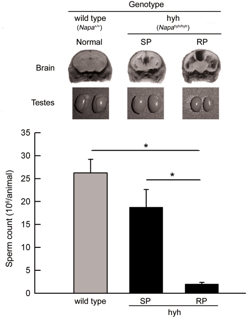
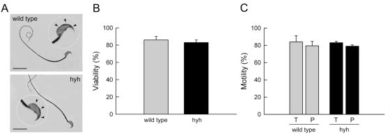
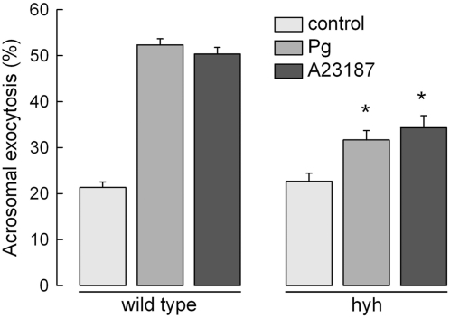
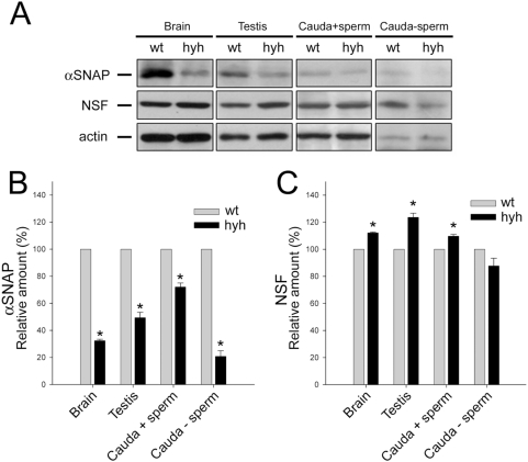
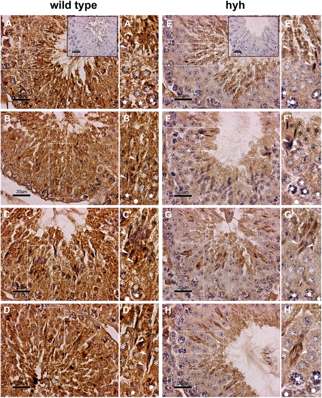
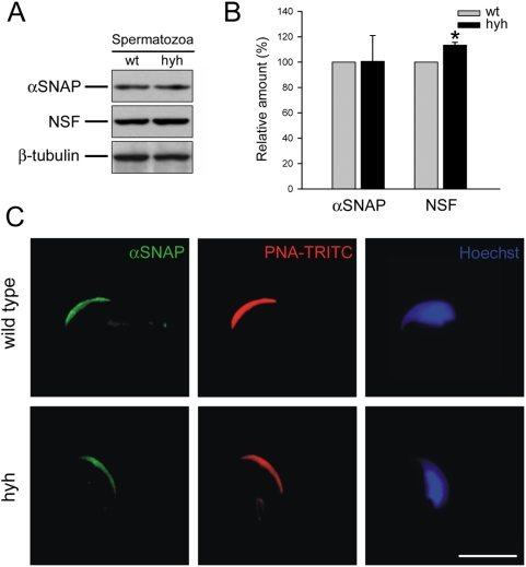
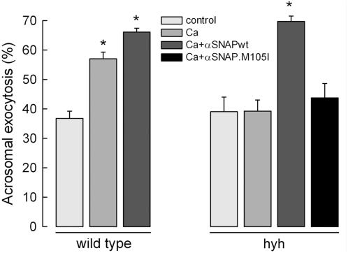
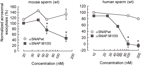
Hydrocephalus with hop gait (hyh) is a recessive inheritable disease that arose spontaneously in a mouse strain. A missense mutation in the Napa gene that results in the substitution of a methionine for isoleucine at position 105 (M105I) of alphaSNAP has been detected in these animals. alphaSNAP is a ubiquitous protein that plays a key role in membrane fusion and exocytosis. In this study, we found that male hyh mice with a mild phenotype produced morphologically normal and motile sperm, but had a strongly reduced fertility. When stimulated with progesterone or A23187 (a calcium ionophore), sperm from these animals had a defective acrosome reaction. It has been reported that the M105I mutation affects the expression but not the function of the protein. Consistent with an hypomorphic phenotype, the testes and epididymides of hyh mice had low amounts of the mutated protein. In contrast, sperm had alphaSNAP levels indistinguishable from those found in wild type cells, suggesting that the mutated protein is not fully functional for acrosomal exocytosis. Corroborating this possibility, addition of recombinant wild type alphaSNAP rescued exocytosis in streptolysin O-permeabilized sperm, while the mutant protein was ineffective. Moreover, addition of recombinant alphaSNAP. M105I inhibited acrosomal exocytosis in permeabilized human and wild type mouse sperm. We conclude that the M105I mutation affects the expression and also the function of alphaSNAP, and that a fully functional alphaSNAP is necessary for acrosomal exocytosis, a key event in fertilization.


Conflict of interest statement

Competing Interests: The authors have declared that no competing interests exist.

Figures

Figure 1
Figure 1. Testes phenotype and sperm count in wild type and hyh (hyh) mice.
Hyh mice from the RP group developed a very severe hydrocephalus. On the other hand, mutant mice from the SP group developed a mild hydrocephalic process and less neuropathological alterations. Brains from wild type (normal phenotype), and SP hyh and RP hyh mice from the same litter (70 day old) were fixed with Bouin solution by vascular perfusion and coronally sectioned at the level of the optic chiasm. Representative images of brain and cerebral ventricles phenotypes are shown. Images of the testes from the same animals are shown below. Cauda epididymidis sperm were obtained as explain in Materials and Methods and counted. Data represent the mean±SEM from at least 6 independent experiments. * p<0.01 (one way ANOVA, Tukey's test).
Figure 2
Figure 2. Sperm characteristics in wild type and SP hyh (hyh) mice.
(A) Bright field micrographs of sperm stained with Coomassie G-250 from wild type and mutant (hyh) mice showing normal cell and acrosome morphology. Scale bar, 15 µm. (B) Sperm viability for wild type and mutant (hyh) mice was assessed by Eosin Y staining. (C) The percentage of motile sperm (T) and having progressive motility (P) was assessed in wild type and mutant (hyh) mice. Data represent the mean±SEM of at least four independent experiments.
Figure 3
Figure 3. Sperm from SP hyh mice have a deficient acrosomal reaction.
Sperm from wild type and SP hyh (hyh) mice were collected from the cauda epididymidis, incubated under capacitation conditions for 1 h and stimulated with buffer (control), 10 µM progesterone (Pg) or 10 µM A23187 (A23187) for 15 min at 37°C. The cells were spotted on slides and fixed in ice-cold methanol. Acrosomal status was evaluated in at least 200 sperm by staining with TRITC-PNA. The data represent the mean±SEM of three independent experiments (*, significant differences between same groups for wild type and hyh mice, P<0.001, Student's t test).
Figure 4
Figure 4. αSNAP and NSF expression in the reproductive tract of wild type (wt) and SP hyh (hyh) mice.
(A) Proteins extracted from testis and cauda epididymidis of wt and SP hyh mice were analyzed by Western blot using an antibody recognizing αSNAP (upper panel) or NSF (middle panel). Signals detected with an anti-actin antibody served as internal controls for equal protein loading (lower panel). Cauda epididymidis extracts were obtained before (Cauda+sperm) and after (Cauda-sperm) sperm were washed out from the organ. Brain was used as a positive control. Blots are representative of 3 or 4 independent experiments. (B, C) Densitometric analysis of Western blot for αSNAP (B) and NSF (C). Black bars (mean±SEM, N = 3 or 4) refer to the relative amount of each protein in hyh samples compared to wt. * p<0.05 (Student's t-test).
Figure 5
Figure 5. Immunolocalization of αSNAP in seminiferous epithelium.
Light micrographs of testis sections from wild type (A–D) and SP hyh mice (E–H) showing general and specific patterns of immunoperoxidase staining using an αSNAP-specific antibody. Background staining with hematoxylin. Different stages of epithelial maturation cycle in normal and mutant testis are presented in comparative mode. A'–H': High magnification images of the regions boxed in the corresponding panel (A–H). Epithelial polarization is oriented upwards. The symbols used are: spermatogonium (white pentagon); primary spermatocytes (white stars); round spermatids (white asterisk); elongated spermatids with high polarization, indicating the residual body or axonemal region (black asterisk) and the heads (white arrowheads). Controls without primary antibodies are shown in the inserts (A and E). In wild type animals, a strong immunoreaction was observed in the whole ephitelium. In contrast, mutant mice showed a notably diminished immunoreaction compared to that of wild type mice (compare E–H to A–D panels). This difference was not evident in spermatids undergoing elongation process. Scale bar, 20 µm.
Figure 6
Figure 6. αSNAP expression and localization in sperm from wild type (wt) and SP hyh (hyh) mice.
(A) Sperm protein extracts obtained from wt and hyh mice were analyzed by Western blot using an antibody recognizing αSNAP (upper panel) or NSF (middle panel). Signals detected with an anti β-tubulin antibody served as internal controls for equal protein loading (lower panel). Blots are representative of seven independent experiments. (B) Densitometric analysis of Western blot for αSNAP and NSF. Black bars (mean±SEM, N = 7) refer to the relative amount of each protein in hyh samples compared to wt (gray bars). * p<0.05 (Student's t-test). (C) αSNAP localizes to the acrosomal region in mouse spermatozoa. Sperm from wild type and hyh mice were fixed, permeabilized and triple-stained with an anti-α/βSNAP antibody (green); TRITC-PNA, a lectin that recognizes the intra-acrosomal content (red); and Hoechst 33258 to visualize the nucleus of the cell (blue). Shown are epifluorescence micrographs of typically stained cells. Scale bar, 10 µm.
Figure 7
Figure 7. αSNAP rescues acrosomal exocytosis in sperm from SP hyh mice.
Sperm from wild type and SP hyh mice were collected from the cauda epididymidis and incubated under capacitation conditions for 1 h. The cells were then permeabilized and stimulated with buffer (control) or 0.5 mM CaCl2 (10 µM free Ca2+). When indicated, 15 nM recombinant wild type αSNAP (αSNAPwt) or the M105I mutant (αSNAP.M105I) were added to the assay. The samples were incubated for 15 min at 37°C. At the end of the incubation, the cells were spotted on slides and fixed in ice-cold methanol. Acrosomal status was evaluated in at least 200 sperm by staining with TRITC-PNA. The data represent the mean±SEM of three independent experiments (*, significant differences with respect to control for wild type and hyh mice, P<0.01, one way ANOVA, Dunnett's test).
Figure 8
Figure 8. Inhibitory effect of αSNAP.M105I on the acrosomal exocytosis of permeabilized normal mouse and human sperm.
(A) Sperm from wild type mice were collected from the cauda epididymidis and incubated under capacitating conditions for 1 h. (B) Human sperm were collected from ejaculates as previously described . Mouse and human cells were then permeabilized and stimulated with 10 µM free Ca2+ in the presence of different concentrations of wild type αSNAP (αSNAPwt) or the M105I mutant (αSNAP.M105I) for 15 min at 37°C. At the end of the incubation, the cells were spotted on slides and fixed in ice-cold methanol. Acrosomal status was evaluated in at least 200 sperm by staining with TRITC-PNA (mouse sperm) or FITC-PSA (human sperm). For each experiment, acrosomal exocytosis was normalized by subtracting the number of reacted spermatozoa in the non-stimulated samples (mean±SEM: 44.8%±4.3% and 11.7%±1.2%, for mouse and human sperm, respectively) from all values, and expressing the resulting values as a percentage of the acrosome reaction observed in cells stimulated with 10 µM free Ca2+ in the absence of added αSNAP (63.4%±3.7% and 23.7%±1.2%, for mouse and human sperm, respectively). The data represent the mean±SEM of three independent experiments. (*, significant differences with respect to wild type αSNAP, P<0.01, Student's t test).

Similar articles

Cited by

References

    1. Whiteheart SW, Griff IC, Brunner M, Clary DO, Mayer T, et al. SNAP family of NSF attachment proteins includes a brain-specific isoform. Nature. 1993;362:353–355. - PubMed
    1. Jahn R, Scheller RH. SNAREs–engines for membrane fusion. Nat Rev Mol Cell Biol. 2006;7:631–643. - PubMed
    1. Wickner W, Schekman R. Membrane fusion. Nat Struct Mol Biol. 2008;15:658–664. - PMC - PubMed
    1. Xu T, Ashery U, Burgoyne RD, Neher E. Early requirement for alpha-SNAP and NSF in the secretory cascade in chromaffin cells. EMBO J. 1999;18:3293–3304. - PMC - PubMed
    1. Morgan A, Burgoyne RD. A role for soluble NSF attachment proteins (SNAPs) in regulated exocytosis in adrenal chromaffin cells. EMBO J. 1995;14:232–239. - PMC - PubMed

Publication types

Substances