. 2008 Mar;6(1):1-20.
doi: 10.2174/157015908783769653.

GABA(A) receptors in normal development and seizures: friends or foes?

Affiliations

GABA(A) receptors in normal development and seizures: friends or foes?

Aristea S Galanopoulou. Curr Neuropharmacol. 2008 Mar.

Abstract

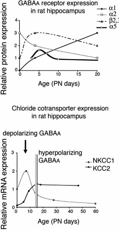
GABA(A) receptors have an age-adapted function in the brain. During early development, they mediate excitatory effects resulting in activation of calcium sensitive signaling processes that are important for the differentiation of the brain. In more mature stages of development and in adults, GABA(A) receptors transmit inhibitory signals. The maturation of GABA(A) signaling follows sex-specific patterns, which appear to also be important for the sexual differentiation of the brain. The inhibitory effects of GABA(A) receptor activation have been widely exploited in the treatment of conditions where neuronal silencing is necessary. For instance, drugs that target GABA(A) receptors are the mainstay of treatment of seizures. Recent evidence suggests however that the physiology and function of GABA(A) receptors changes in the brain of a subject that has epilepsy or status epilepticus.This review will summarize the physiology of and the developmental factors regulating the signaling and function of GABA(A) receptors; how these may change in the brain that has experienced prior seizures; what are the implications for the age and sex specific treatment of seizures and status epilepticus. Finally, the implications of these changes for the treatment of certain forms of medically refractory epilepsies and status epilepticus will be discussed.

Keywords: GABA; brain; chloride; development; expression; hippocampus; physiology.; seizure.


Figures

Fig. (1).
Fig. (1).
Developmental changes in selected GABAA receptor subunits, chloride cotransporters (KCC2 and NKCC1) and GABAA receptor physiology in rat hippocampus.Upper panel: A developmental decrease in α2 and α5 in parallel with an increase in a1 has been described in rat hippocampus. Age dependent changes in other subunits, such as β2,3 has also been reported. Results are from studies [34, 74, 89, 120, 140, 253, 204]. The scale is arbitrary and intends to depict relative changes in expression of a given subunit across ages, and not the relative abundance of one subunit vs another. Lower panel: A switch from an NKCC1-dominant to a KCC2-dominant state occurs in infantile rat hippocampus and has been implicated in the functional switch of GABAA receptor signaling from depolarizing to hyperpolarizing. Results are compiled from studies in male rats or rats of undetermined sex [247, 261]. The vertical bar indicates the age when hyperpolarizing GABAA receptor signaling occurs.
Fig. (2)
Fig. (2)
The developmental switch in chloride cotransporter (CCC) expression drives the functional switch of GABAA receptors from depolarizing to hyperpolarizing.The developmental increase in KCC2 and, in certain tissues, the decrease in NKCC1 triggers the switch from depolarizing to hyperpolarizing GABAAergic signaling [247, 261]. GABA-mediated depolarizations activate L-type voltage sensitive calcium channels(L-VSCC) and release the Mg++ block of NMDA receptors, increasing intracellular Ca++. This can activate calciumregulated signaling pathways, which are important in neuronal development, migration, proliferation, synaptogenesis and differentiation. The GABA-mediated activation of calcium signaling does not occur in neurons with hyperpolarizing GABAA receptor responses.
Fig. (3) Schematic depiction of selected proteins involved in the regulation of Cl- homeostasis.
Upper panel: Cl- accumulation is effected by the presence of the Na+/Cl- Cotransporter NCC and Na+/K+/Cl- Cotransporter NKCCs, with main representative being NKCC1. Their function is dependent upon the supply of Na+ by the Na+/K+ ATPase. In contrast, Anion Exchangers, like AE3, favor Cl- accumulation in a sodium independent manner. Lower panel: Low intracellular Cl- concentration occurs as a result of K+/Cl- Cotransporters, such as KCC2, which export Cl- and K+. As a result, their function is also dependent upon K+ supply by Na+/K+ ATPases. The Sodium Dependent Anion Exchangers NDAE and Chloride Channel 2 (Clc2) also decrease intracellular Cl-[69, 268].
Fig. (4)
Fig. (4)
Differential regulation of KCC2 in neurons with depolarizing or hyperpolarizing GABAAergic signaling.GABAA receptor activation and BDNF increase KCC2 in immature neurons with depolarizing GABAA ergic responses, but decrease it in neurons with hyperpolarizing GABAA ergic signaling. Estradiol (E) downregulates KCC2 only in neurons with depolarizing GABA. Testosterone and its androgenic products (T) increase KCC2 in both conditions [3, 35, 81, 262, 263, 317].
Fig. (5)Schematic depiction of the timeline of changes in GABA<sub>A</sub> receptor subunit mRNA expression in the hippocampus, in rodent models of temporal lobe seizures and epilepsy.
Fig. (5)Schematic depiction of the timeline of changes in GABAA receptor subunit mRNA expression in the hippocampus, in rodent models of temporal lobe seizures and epilepsy.
The effects of SE change according to age of induction, model, and species. In most cases, the results stem from the lithium-pilocarpine or pilocarpine SE model, except for the results marked with an asterisk, which were described after kainic acid SE. Adulthood starts at PN60. The time scale used for the effects of SE in adult rats is approximate and is meant to reflect changes during the latent phase of epileptogene-sis, prior to the onset of spontaneous seizures, and during the epileptic phase, ie after the occurrence of 2 spontaneous seizures. The diagrams are based on a review of the pertinent literature [33, 73, 163, 254, 244, 345].

References

    1. Adelson PD, Black PM, Madsen JR, Kramer U, Rockoff MA, Riviello JJ, Helmers SL, Mikati M, Holmes GL. Use of subdural grids and strip electrodes to identify a seizure focus in children. Pediatr. Neurosurg. 1995;22(4):174–80. - PubMed
    1. Adkins CE, Pillai GV, Kerby J, Bonnert TP, Haldon C, McKernan RM, Gonzalez JE, Oades K, Whiting PJ, Simpson PB. Alpha4beta3delta GABA(A) receptors characterized by fluorescence resonance energy transfer-derived measurements of membrane potential. J. Biol. Chem. 2001;276(42):38934–9. - PubMed
    1. Aguado F, Carmona MA, Pozas E, Aguilo A, Martinez-Guijarro FJ, Alcantara S, Borrell V, Yuste R, Ibanez CF, Soriano E. BDNF regulates spontaneous correlated activity at early developmental stages by increasing synaptogenesis and expression of the K+/Cl- co-transporter KCC2. Development. 2003;130(7):1267–80. - PubMed
    1. Aickin CC, Deisz RA, Lux HD. Mechanisms of chloride transport in crayfish stretch receptor neurones and guinea pig vas deferens: implications for inhibition mediated by GABA. Neurosci. Lett. 1984;47(3):239–44. - PubMed
    1. Akaike N, Inomata N, Yakushiji T. Differential effects of extra- and intracellular anions on GABA-activated currents in bullfrog sensory neurons. J. Neurophysiol. 1989;62(6):1388–99. - PubMed

LinkOut - more resources