

Neurodegenerative diseases of the retina and potential for protection and recovery
- PMID: 19305795
- PMCID: PMC2647152
- DOI: 10.2174/157015908784533851
Neurodegenerative diseases of the retina and potential for protection and recovery
Abstract
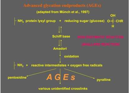
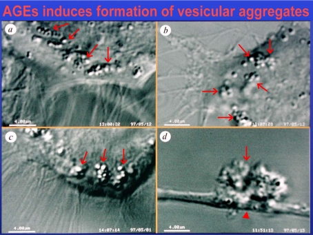
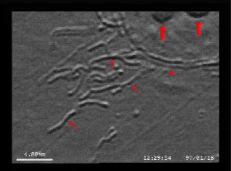
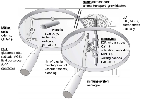
Recent advances in our understanding of the mechanisms in the cascade of events resulting in retinal cell death in ocular pathologies like glaucoma, diabetic retinopathy and age-related macular degeneration led to the common descriptive term of neurodegenerative diseases of the retina. The final common pathophysiologic pathway of these diseases includes a particular form of metabolic stress, resulting in an insufficient supply of nutrients to the respective target structures (optic nerve head, retina). During metabolic stress, glutamate is released initiating the death of neurones containing ionotropic glutamate (N-methyl-D-aspartat, NMDA) receptors present on ganglion cells and a specific type of amacrine cells. Experimental studies demonstrate that several drugs reduce or prevent the death of retinal neurones deficient of nutrients. These agents generally block NMDA receptors to prevent the action of glutamate or halt the subsequent pathophysiologic cycle resulting in cell death. The major causes for cell death following activation of NMDA receptors are the influx of calcium and sodium into cells, the generation of free radicals linked to the formation of advanced glycation endproducts (AGEs) and/or advanced lipoxidation endproducts (ALEs) as well as defects in the mitochondrial respiratory chain. Substances preventing these cytotoxic events are considered to be potentially neuroprotective.
Keywords: Neurodegeneration; age-related macular degeneration; diabetic retinopathy; glaucoma; neuroprotection; retina; retinal ganglion cells..
Figures
References
-
- Ahmed N, Ahmed U, Thornalley PJ, Hager K, Fleischer G, Munch G. Protein Glycation, Oxidation and Nitration Adduct Residues and Free Adducts of Cerebrospinal Fluid in Alzheimer's Disease and Link to Cognitive Impairment. J. Neurochem. 2005;92:255–263. - PubMed
-
- Alizadeh M, Wada M, Gelfman CM, Handa JT, Hjelmeland LM. Downregulation of Differentiation Specific Gene Expression by Oxidative Stress in ARPE-19 cells. Invest. Ophthalmol. Vis. Sci. 2001;42:2706–2713. - PubMed
-
- Anderson DR. Glaucoma, Capillaries and Pericytes. 1. Blood Flow Regulation. Ophthalmologica. 1996;210:257–262. - PubMed
-
- Anderson DR, Davis EB. Glaucoma, Capillaries and Pericytes. 5. Preliminary Evidence that Carbon Dioxide Relaxes Pericyte Contractile Tone. Ophthalmologica. 1996;210:280–284. - PubMed
-
- Anderson DR, Davis EB. Glaucoma, Capillaries and Pericytes. 2. Identification and Characterization of Retinal Pericytes in Culture. Ophthalmologica. 1996;210:263–268. - PubMed
LinkOut - more resources
Full Text Sources
Other Literature Sources