. 2009 Jul;47(7):719-29.
doi: 10.1007/s11517-009-0472-x. Epub 2009 Mar 21.

Cardiac anisotropy in boundary-element models for the electrocardiogram

Affiliations

Cardiac anisotropy in boundary-element models for the electrocardiogram

Mark Potse et al. Med Biol Eng Comput. 2009 Jul.

Abstract

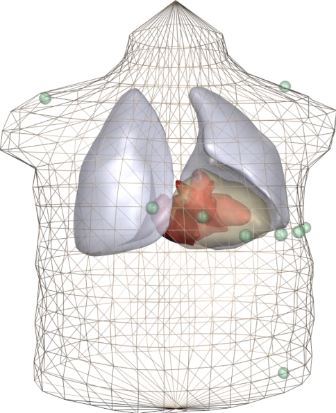
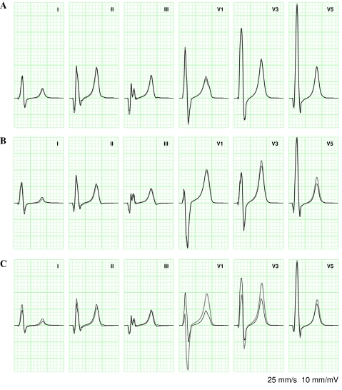
The boundary-element method (BEM) is widely used for electrocardiogram (ECG) simulation. Its major disadvantage is its perceived inability to deal with the anisotropic electric conductivity of the myocardial interstitium, which led researchers to represent only intracellular anisotropy or neglect anisotropy altogether. We computed ECGs with a BEM model based on dipole sources that accounted for a "compound" anisotropy ratio. The ECGs were compared with those computed by a finite-difference model, in which intracellular and interstitial anisotropy could be represented without compromise. For a given set of conductivities, we always found a compound anisotropy value that led to acceptable differences between BEM and finite-difference results. In contrast, a fully isotropic model produced unacceptably large differences. A model that accounted only for intracellular anisotropy showed intermediate performance. We conclude that using a compound anisotropy ratio allows BEM-based ECG models to more accurately represent both anisotropies.


Figures

Fig. 1
Fig. 1
Anatomic model. The triangulation of the torso corresponds to that used in the BEM model. For clarity, other components are shown as smooth surfaces. The standard ECG electrodes (three limb electrodes and six precordial electrodes) are shown as green spheres. For actual simulations the torso surface was replaced by inner and outer surfaces of the skeletal muscle layer, and electrodes moved to the outer layer
Fig. 2
Fig. 2
Comparison of ECGs simulated with an FD model (gray) and with a BEM model (black). ECGs were obtained from a normal (sinus rhythm) activation sequence. A representative subset of the standard 12-lead ECG is shown. ECGs are displayed in the conventional way, using grid lines with 40 ms spacing horizontally and 0.1 mV spacing vertically, and no axis labels. a Both models isotropic (RD = 0.08). b Both models anisotropic (RD = 0.13, Table 3). c Fully isotropic BEM versus anisotropic FD model (RD = 0.59, Table 4)

Similar articles

Cited by

References

    1. {'text': '', 'ref_index': 1, 'ids': [{'type': 'DOI', 'value': '10.1109/TBME.1987.326079', 'is_inner': False, 'url': 'https://doi.org/10.1109/tbme.1987.326079'}, {'type': 'PubMed', 'value': '3610193', 'is_inner': True, 'url': 'https://pubmed.ncbi.nlm.nih.gov/3610193/'}]}
    2. Aoki M, Okamoto Y, Musha T, Harumi KI (1987) Three-dimensional simulation of the ventricular depolarization and repolarization processes and body surface potentials: normal heart and bundle branch block. IEEE Trans Biomed Eng 34(6):454–462 - PubMed
    1. {'text': '', 'ref_index': 1, 'ids': [{'type': 'DOI', 'value': '10.1016/S0006-3495(67)86598-6', 'is_inner': False, 'url': 'https://doi.org/10.1016/s0006-3495(67)86598-6'}, {'type': 'PMC', 'value': 'PMC1368073', 'is_inner': False, 'url': 'https://pmc.ncbi.nlm.nih.gov/articles/PMC1368073/'}, {'type': 'PubMed', 'value': '6048873', 'is_inner': True, 'url': 'https://pubmed.ncbi.nlm.nih.gov/6048873/'}]}
    2. Barnard AC, Duck IM, Lynn MS (1967) The application of electromagnetic theory to electrocardiology; I. Derivation of the integral equations. Biophys J 7:443–462 - PMC - PubMed
    1. {'text': '', 'ref_index': 1, 'ids': [{'type': 'DOI', 'value': '10.1016/S0006-3495(67)86599-8', 'is_inner': False, 'url': 'https://doi.org/10.1016/s0006-3495(67)86599-8'}, {'type': 'PMC', 'value': 'PMC1368074', 'is_inner': False, 'url': 'https://pmc.ncbi.nlm.nih.gov/articles/PMC1368074/'}, {'type': 'PubMed', 'value': '6058137', 'is_inner': True, 'url': 'https://pubmed.ncbi.nlm.nih.gov/6058137/'}]}
    2. Barnard AC, Duck IM, Lynn MS, Timlake WP (1967) The application of electromagnetic theory to electrocardiology; II. Numerical solution of the integral equations. Biophys J 7:463–490 - PMC - PubMed
    1. {'text': '', 'ref_index': 1, 'ids': [{'type': 'DOI', 'value': '10.1109/TBME.1966.4502411', 'is_inner': False, 'url': 'https://doi.org/10.1109/tbme.1966.4502411'}, {'type': 'PubMed', 'value': '5964789', 'is_inner': True, 'url': 'https://pubmed.ncbi.nlm.nih.gov/5964789/'}]}
    2. Barr RC, Pilkington TC, Boineau JP, Spach MS (1966) Determining surface potentials from current dipoles, with application to electrocardiography. IEEE Trans Biomed Eng 13(2):88–92 - PubMed
    1. {'text': '', 'ref_index': 1, 'ids': [{'type': 'DOI', 'value': '10.1114/1.1527045', 'is_inner': False, 'url': 'https://doi.org/10.1114/1.1527045'}, {'type': 'PubMed', 'value': '12540206', 'is_inner': True, 'url': 'https://pubmed.ncbi.nlm.nih.gov/12540206/'}]}
    2. Buist M, Pullan A (2002) Torso coupling techniques for the forward problem of electrophysiology. Ann Biomed Eng 30:1299–1312 - PubMed

LinkOut - more resources