. 2009 Sep;76(4):882-94.
doi: 10.1002/prot.22394.

Construct optimization for protein NMR structure analysis using amide hydrogen/deuterium exchange mass spectrometry

Affiliations

Construct optimization for protein NMR structure analysis using amide hydrogen/deuterium exchange mass spectrometry

Seema Sharma et al. Proteins. 2009 Sep.

Abstract

Disordered or unstructured regions of proteins, while often very important biologically, can pose significant challenges for resonance assignment and three-dimensional structure determination of the ordered regions of proteins by NMR methods. In this article, we demonstrate the application of (1)H/(2)H exchange mass spectrometry (DXMS) for the rapid identification of disordered segments of proteins and design of protein constructs that are more suitable for structural analysis by NMR. In this benchmark study, DXMS is applied to five NMR protein targets chosen from the Northeast Structural Genomics project. These data were then used to design optimized constructs for three partially disordered proteins. Truncated proteins obtained by deletion of disordered N- and C-terminal tails were evaluated using (1)H-(15)N HSQC and (1)H-(15)N heteronuclear NOE NMR experiments to assess their structural integrity. These constructs provide significantly improved NMR spectra, with minimal structural perturbations to the ordered regions of the protein structure. As a representative example, we compare the solution structures of the full length and DXMS-based truncated construct for a 77-residue partially disordered DUF896 family protein YnzC from Bacillus subtilis, where deletion of the disordered residues (ca. 40% of the protein) does not affect the native structure. In addition, we demonstrate that throughput of the DXMS process can be increased by analyzing mixtures of up to four proteins without reducing the sequence coverage for each protein. Our results demonstrate that DXMS can serve as a central component of a process for optimizing protein constructs for NMR structure determination.


Figures

Figure 1
Figure 1
(A) General strategy for construct optimization of targets for structure determination in the NESG consortium. After initial NMR screening, disorder prediction results for targets exhibiting evidence of partial disorder are classified into three groups: A) consensus, B) multiple disordered regions, and C) poor consensus. Construct optimization for Class A targets is based exclusively on the bioinformatics predictions. Class B and C targets, however, are subsequently analyzed by DXMS so as to accurately determine the ordered/disordered boundaries, and optimized constructs are designed on the basis of these data. Finally, construct-optimized targets are re-evaluated by 1H -15N HSQC NMR and sent for crystallization screening. (B) General protocol for DXMS analysis of protein targets in the NESG consortium. The major steps in the protocol are as follows: mixing the protein sample(s) with 2H2O (depicted on the right with yellow circles), quenching the 1H/2H exchange at specific time points by lowering the pH, pepsin digestion, and separation of peptide fragments by LC-MS. See Material and Methods section for a complete description of the DXMS protocol used in this work.
Figure 2
Figure 2
Overlay of 1H -15N HSQC NMR spectra (25 °C) of TPPP family protein C32E8.3 from C. elegans (NESG target WR33) for the full length (1 - 180) protein (blue), and truncated (1 - 115) protein construct (red). Side chain Arg peaks aliased in the 15N-dimension of the spectrum of truncated C32E8.3 are boxed.
Figure 3
Figure 3
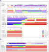
Protein sequence, NMR secondary structure, 1H-15N hetNOE, and DXMS results (10, 100, and 1000 second exchange durations; pH 7.5 and temperature ~ 0 °C) analyzed individually (I) and in a four protein mixture (M) for (A) protein C32E8.3 from C. elegans (NESG target WR33) and (B) protein YnzC from B. subtilis (NESG target SR384).
Figure 4
Figure 4
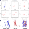
(A) 1H-15N HSQC NMR spectra (20 °C) for the full length (1 - 77) protein YnzC from B. subtilis (NESG target SR384) and five truncated protein constructs that were designed based on the DXMS results. (B) NMR solution structures of full length (blue; PDB ID, 2HEP) and truncated (red; PDB ID, 2JVD) protein YnzC from B. subtilis (NESG targets SR384 and SR384-1-46, respectively). The first two images show ribbon diagrams of representative (lowest energy) conformers of full length and truncated YnzC. The superimposed final ensembles of structures (rotated 180°) are presented on the right (20 models each; heavy atoms for residues 2 to 40 are shown). The backbone RMSD between the mean coordinates of the ordered residues encompassing the helices (5-19 and 22-38) of each ensemble is 0.84 Å.
Figure 5
Figure 5
DXMS-based construct optimization of E. coli yiaD (NESG target, ER553). (A) 1H-15N HSQC spectra (20 °C) of full length (left) and construct optimized (right) E. coli yiaD (59-199). (B) DXMS results for full length E. coli yiaD (10, 100, and 1000 second exchange durations at pH 7.5 and ~ 0 °C). The PROF secondary structure prediction results are shown above the DXMS data.
Figure 6
Figure 6
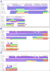
(A) Protein sequence, experimentally-determined secondary structure, 1H-15N hetNOE, and DXMS results (10, 100, and 1000 second exchange durations at pH 7.5 and ~ 0 °C) for cytoplasmic protein Q8ZRJ2 (NESG target StR65) analyzed individually (I) and in a four protein mixture (M). (B and C) Protein sequence, experimentally-determined secondary structure, 1H-15N hetNOE, and DXMS results (10, 100, and 1000 second exchange durations) for YjcQ protein from B. subtilis (NESG target SR346). (B) Results at pH 7.5 and ~ 0 °C for the protein analyzed individually (I) and in a four protein mixture (M). (C) Results at pH 5.5 and ~ 0 °C (in 20 mM ammonium acetate, 0.1 M NaCl, 5 mm CaCl2) for the protein analyzed individually (I).

References

    1. Cavanagh J, Fairbrother WJ, Palmer AG, III, Skelton NJ, Rance M. Protein NMR Spectroscopy: Principles and Practice. 2. New York: Elsevier Academic Press; 2007. p. 912.
    1. Kay LE. NMR studies of protein structure and dynamics. J Magn Reson. 2005;173:193–207. - PubMed
    1. Bax A, Grishaev A. Weak alignment NMR: A hawk-eyed view of biomolecular structure and dynamics. Curr Opin Struct Biol. 2005;15:563–570. - PubMed
    1. Liu J, Montelione GT, Rost B. Novel leverage of structural genomics. Nature Biotechnol. 2007;25:849–851. - PubMed
    1. Dyson HJ, Wright PE. Intrinsically unstructured proteins and their functions. Nature Rev. 2005;6:197–208. - PubMed

Publication types

MeSH terms