. 2009 Jun 1;237(2):137-45.
doi: 10.1016/j.taap.2009.03.006. Epub 2009 Mar 21.

Satratoxin G interaction with 40S and 60S ribosomal subunits precedes apoptosis in the macrophage

Affiliations

Satratoxin G interaction with 40S and 60S ribosomal subunits precedes apoptosis in the macrophage

Hee Kyong Bae et al. Toxicol Appl Pharmacol. .

Abstract

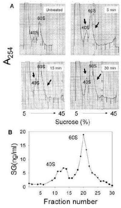
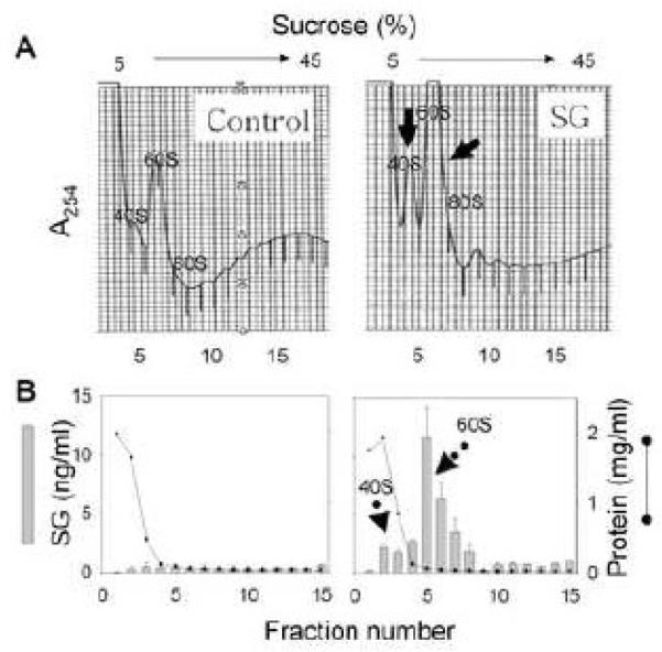
Satratoxin G (SG) and other macrocyclic trichothecene mycotoxins are potent inhibitors of eukaryotic translation that are potentially immunosuppressive. The purpose of this research was to test the hypothesis that SG-induced apoptosis in the macrophage correlates with binding of this toxin to the ribosome. Exposure of RAW 264.7 murine macrophages to SG at concentrations of 10 to 80 ng/ml induced DNA fragmentation within 4 h that was indicative of apoptosis. To relate these findings to ribosome binding of SG, RAW cells were exposed to different toxin concentrations for various time intervals, ribosomal fractions isolated by sucrose density gradient ultracentrifugation and resultant fractions analyzed for SG by competitive ELISA. SG was found to specifically interact with 40S and 60S ribosomal subunits as early as 5 min and that, at high concentrations or extended incubation times, the toxin induced polysome disaggregation. While co-incubation with the simple Type B trichothecene DON had no effect on SG uptake into cell cytoplasm, it inhibited SG binding to the ribosome, suggesting that the two toxins bound to identical sites and that SG binding was reversible. Although both SG and DON induced mobilization of p38 and JNK 1/2 to the ribosome, phosphorylation of ribosomal bound MAPKs occurred only after DON treatment. SG association with the 40S and 60S subunits was also observed in the PC-12 neuronal cell model which is similarly susceptible to apoptosis. To summarize, SG rapidly binds small and large ribosomal subunits in a concentration- and time-dependent manner that was consistent with induction of apoptosis.


Figures

Figure 1
Figure 1. SG induces apoptosis in RAW 264.7 cells
Cells were treated with different SG concentrations for various time intervals and then assessed for DNA fragmentation by (A) cell-death ELISA in duplicate or (B) agarose gel electrophoresis.
Figure 2
Figure 2. UV absorption scan for SG
Purified SG was diluted in phosphate buffer (pH 7.0) and absorbance measured over the UV spectrum.
Figure 3
Figure 3. SG binding to ribosomal subunits is concentration-dependent in RAW 264.7 cells
Cells were treated with SG (0, 20, 40, 80 ng/ml) for 1 h and then lysed with polysome extraction buffer (PEB). (A) Ribosomal fractions were separated on a sucrose gradient and the A254 monitored and small subunit, large subunit, monosome and ploysomes labeled as 40S, 60S, 80S and P, respectively, (B) Ribosomal subunits and monosome (RS+M) fractions were pooled and analyzed for SG by competitive ELISA and for total protein colorimetrically.
Figure 4
Figure 4. Kinetics of SG binding to ribosomal in RAW 264.7 cells
Cells were untreated or treated (A) with water or SG (10 ng/ml) for 15 (B), 30 (C), 60 (D), 120 (E), and 240 (F) min and then lysed with PEB. Ribosomal fractions were separated on a sucrose gradient A254 and monitored. Individual fractions (0.5 ml) were analyzed for SG (open circle) and total protein (vertical bar). Solid arrows indicate increased absorbances with increasing SG concentration. Dotted arrows indicate increased SG is detectable in ribosomal fractions as toxin concentration ins increased.
Figure 5
Figure 5. High SG concentration induces rapid, saturable binding to 40S and 60S ribosomal subunits in RAW 264.7 cells
(A)Cells were untreated or treated with SG (100 ng/ml) for 5, 15, 30 min and then lysed with PEB. Ribosomal fractions were separated on a sucrose gradient system and A254 monitored. (B) Cells were treated with SG (100 ng/ml) for 15 min. PEB lysate were separated at high resolution (0.25 ml/fraction) on a sucrose gradient and then fractions analyzed by SG ELISA. Comparable treatment with water vehicle only revealed no detectable SG (data not shown)
Figure 6
Figure 6. DON competitively inhibits SG binding to 40S and 60S ribosomal subunits in RAW 264.7 cells
Cells were treated with vehicle, SG (100 ng/ml), DON (500 ng/ml) or both toxins for 15 min and then lysed with PEB. (A) SG content of cell lysate was analyzed by ELISA. (B) Ribosomal fractions were separated on a sucrose gradient and A254 monitored.
Figure 7
Figure 7. SG binding to the ribosome is reversible in RAW 264.7 cells
Cells were incubated with SG (20 ng/ml) for 60 min and ribosomal fractions were separated on sucrose gradient. Pooled RS+M fractions , all containing bound SG, were incubated with additional SG (20 ng/ml) and/or (DON 500 ng/ml) for 1 h at 37°C. RS+M were repeatedly concentrated and washed to remove free SG and then analyzed by competitive SG ELISA.
Figure 8
Figure 8. SG induces modest p38 and JNK phosphorylation in RAW 264.7 cells
Cells were treated with SG (100-500 ng/ml) or DON (100 ng/ml) for 30 min and then lysed with SDS buffer. Total protein was analyzed by Western blotting with specific antibodies to non-phosphorylated p38 and JNK and their phosphorylated forms.
Figure 9
Figure 9. SG and DON induce MAPK interaction with the ribosome
Cells were treated with vehicle, SG (100 ng/ml), DON (500 ng/ml) or both toxins for 15 min, and then lysed with PEB. (A) whole cell lysates were analyzed by Western blotting using specific antibodies to phosphorylated p38 and JNK and their non-phosphorylated forms. (B) Lysates were fractionated on sucrose gradient and pooled ribosomal subunits and monosomes (RS+M) analyzed by Western blotting.
Figure 10
Figure 10. SG binds to 40S and 60S ribosomal protein fractions in PC-12 neuronal cells
Cells were treated with water or SG (10 ng/ml) for 60 min and lysed with PEB. Ribosomal fractions were separated on a sucrose gradient system and A254 monitored. Individual fractions were analyzed for SG and protein.

References

    1. Bae HK, Pestka JJ. Deoxynivalenol induces p38 interaction with the ribosome in Monocytes and Macrophages. Toxicol. Sci. 2008;105:59–66. - PMC - PubMed
    1. Bamburg JR. Biological and biochemical actions of trichothecene mycotoxins. Prog. Mol. Subcell. Biol. 1983;8:41–110.
    1. Carter CJ, Cannon M. Structural requirements for the inhibitory action of 12,13-epoxytrichothecenes on protein synthesis in eukaryotes. Biochem. J. 1977;166:399–409. - PMC - PubMed
    1. Chung YJ, Jarvis BB, Tak H, Pestka JJ. Immunochemical assay for satratoxin G and other macrocyclic trichothecenes associated with indoor air contamination by Stachybotrys chartarum. Toxicol. Mech. Meth. 2003a;13:247–252. - PubMed
    1. Chung YJ, Zhou HR, Pestka JJ. Transcriptional and posttranscriptional roles for p38 mitogen-activated protein kinase in upregulation of TNF-alpha expression by deoxynivalenol (vomitoxin) Toxicol. Appl. Pharmacol. 2003b;193:188–201. - PubMed

Publication types

MeSH terms