Review
. 2009 May;32(5):249-56.
doi: 10.1016/j.tins.2009.01.006. Epub 2009 Mar 21.

Calcium homeostasis, selective vulnerability and Parkinson's disease

Affiliations
Review

Calcium homeostasis, selective vulnerability and Parkinson's disease

C Savio Chan et al. Trends Neurosci. 2009 May.

Abstract

Parkinson's disease (PD) is a common neurodegenerative disorder of which the core motor symptoms are attributable to the degeneration of dopamine (DA) neurons in the substantia nigra pars compacta (SNc). Recent work has revealed that the engagement of L-type Ca(2+) channels during autonomous pacemaking renders SNc DA neurons susceptible to mitochondrial toxins used to create animal models of PD, indicating that homeostatic Ca(2+) stress could be a determinant of their selective vulnerability. This view is buttressed by the central role of mitochondria and the endoplasmic reticulum (linchpins of current theories about the origins of PD) in Ca(2+) homeostasis. Here, we summarize this evidence and suggest the dual roles had by these organelles could compromise their function, leading to accelerated aging of SNc DA neurons, particularly in the face of genetic or environmental stress. We conclude with a discussion of potential therapeutic strategies for slowing the progression of PD.


Figures
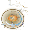
Figure 1
Figure 1
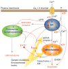
Ca2+ transport in SNc DA neurons. The steep concentration gradient for Ca2+ enables it to cross the plasma membrane readily into cells through open pores such as L-type Ca2+ channels. Once inside neurons, it is either transported back across the plasma membrane or sequestered in intracellular organelles. Ca2+ is transported across the plasma membrane through either the Ca2+-ATPase (PMCA) or through a Na+/Ca2+ exchanger (NCX) that relies upon the Na+ gradient. Ca2+ is rapidly sequestered either by ionic interactions with buffering proteins or by transport into cytosolic organelles (i.e. the mitochondria and the ER). The ER uses high-affinity smooth ER Ca2+ (SERCA) pumps that depend upon ATP to take Ca2+ from the cytoplasm into the ER lumen. Ca2+ flows back into the cytoplasm after the opening of inositol trisphosphate receptors (IP3R) and ryanodine receptors (RyR) studding the ER membrane. Mitochondria are often found in close apposition to the ER and plasma membrane, creating a region of high (but localized) Ca2+ concentration that drives Ca2+ into the matrix of mitochondria through a Ca2+ uniporter. Ca2+ can leave the mitochondrion through a number of mechanisms. The dominant mitochondrial Ca2+-efflux path in neurons is through mitochondrial NCXs. Ca2+ release through higher conductance ion channels, such as the mitochondrial permeability transition pore (mPTP), has also been proposed. The mPTP is posited to have two conductance states: a low-conductance state that is reversible and participates in physiological Ca2+ handling, and a high-conductance state that is irreversible and leads to mitochondrial swelling and loss of molecules such as cytochrome c that trigger apoptosis (Figure 2). Also shown schematically are elements of the tricarboxylic acid (TCA) cycle that produces reducing equivalents for the electron transport chain; complexes I–IV are shown in red. The electrochemical gradient created drives ATP synthase and the conversion of ATP from adenosine diphosphate (ADP) delivered to the matrix by the adenine nucleotide translocator (ANT).
Figure 2
Figure 2
The role played by the ER and mitochondria in Ca2+ homeostasis could contribute to Lewy body (LB) formation and premature death of SNc DA neurons. Ca2+ entry through plasma membrane Cav1.3 Ca2+ channels during activity is either pumped back across the plasma membrane or rapidly sequestered in the ER or mitochondria (Figure 1). Both processes require energy stored in the form of ATP (red ‘ATP’ labels denote ATP requirement). The metabolic demand created by these ATP-dependent steps in Ca2+ homeostasis should increase oxidative phosphorylation in mitochondria and the production of damaging ROS. ROS damage mitochondrial proteins such as complex I and mtDNA, reducing the efficiency of oxidative phosphorylation (negative consequences are symbolized by red circles; positive or augmenting consequences are symbolized by red arrows). In extreme cases, the stress on mitochondria induces mPTP opening, swelling and the release of cytochrome c and other pro-apoptotic proteins such as apoptosis-inducing factor (AIF). In parallel, ROS are capable of damaging ER proteins, elevating the concentration of misfolded proteins that need to be degraded by proteasomes and autophagosomes. The unfolded protein response (UPR) triggered by this elevation in misfolded proteins should further reduce ER production of proteins and potentially lead to the release of pro-apoptotic factors such as C/EBP homologous protein (CHOP). The role of mitochondria in Ca2+ homeostasis could further compromise their ability to generate ATP, leading to a functionally important drop in cytosolic ATP levels. Such a drop would compromise both ER and proteasome and autophagosome function, also promoting the formation of protein aggregates such as LBs. Genetic mutations (Box 1) or environmental toxins such as rotenone could further compromise mitochondrial or ER function, rendering them more vulnerable to Ca2+-induced stress. By hastening the decline in ER and mitochondrial function and the accelerated loss of SNc DA neurons, these genetic and environmental factors could be seen as ‘causing’ PD.

References

    1. de Lau LM, et al. Incidence of parkinsonism and Parkinson disease in a general population: the Rotterdam Study. Neurology. 2004;63:1240–1244. - PubMed
    1. de Rijk MC, et al. Prevalence of parkinsonism and Parkinson’s disease in Europe: the EUROPARKINSON Collaborative Study. European Community Concerted Action on the Epidemiology of Parkinson’s disease. J Neurol Neurosurg Psychiatry. 1997;62:10–15. - PMC - PubMed
    1. Dorsey ER, et al. Projected number of people with Parkinson disease in the most populous nations, 2005 through 2030. Neurology. 2007;68:384–386. - PubMed
    1. Hornykiewicz O. Dopamine (3-hydroxytyramine) and brain function. Pharmacol Rev. 1966;18:925–964. - PubMed
    1. Riederer P, Wuketich S. Time course of nigrostriatal degeneration in parkinson’s disease. A detailed study of influential factors in human brain amine analysis. J Neural Transm. 1976;38:277–301. - PubMed

Publication types