

Proliferative capacity of vein graft smooth muscle cells and fibroblasts in vitro correlates with graft stenosis
- PMID: 19307078
- PMCID: PMC2692862
- DOI: 10.1016/j.jvs.2008.12.020
Proliferative capacity of vein graft smooth muscle cells and fibroblasts in vitro correlates with graft stenosis
Abstract
Objective: About a quarter of peripheral vein grafts fail due in part to intimal hyperplasia. The proliferative capacity and response to growth inhibitors of medial smooth muscle cells and adventitial fibroblasts in vitro were studied to test the hypothesis that intrinsic differences in cells of vein grafts are associated with graft failure.
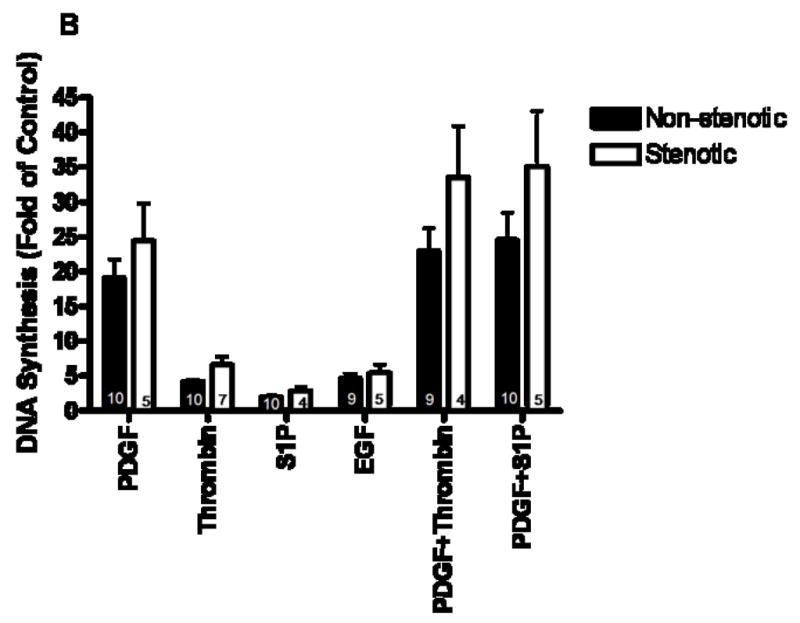
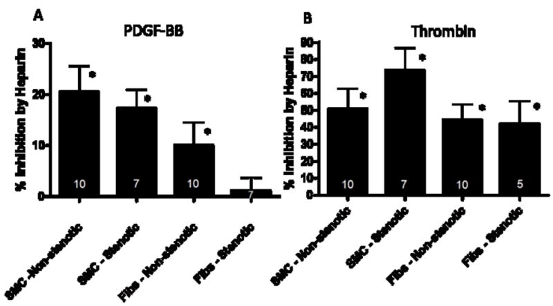
Methods: Cells were grown from explants of the medial and adventitial layers of samples of vein grafts obtained at the time of implantation. Vein graft patency and function were monitored over the first 12 months using ankle pressures and Duplex ultrasound to determine vein graft status. Cells were obtained from veins from 11 patients whose grafts remained patent (non-stenotic) and from seven patients whose grafts developed stenosis. Smooth muscle cells (SMCs) derived from media and fibroblasts derived from adventitia were growth arrested in serum-free medium and then stimulated with 1 muM sphingosine-1-phosphate (S1P), 10 nM thrombin, 10 ng/ml epidermal growth factor (EGF), 10 ng/ml platelet-derived growth factor-BB (PDGF-BB), PDGF-BB plus S1P, or PDGF-BB plus thrombin for determination of incorporation of [(3)H]-thymidine into DNA. Cells receiving PDGF-BB or thrombin were also treated with or without 100 microg/ml heparin, which is a growth inhibitor. Cells receiving thrombin were also treated with or without 150 nM AG1478, an EGF receptor kinase inhibitor.
Results: SMCs and fibroblasts from veins of patients that developed stenosis responded more to the growth factors, such as PDGF-BB alone or in combination with thrombin or S1P, than cells from veins of patients that remained patent (P = .012). In addition, while PDGF-BB-mediated proliferation of fibroblasts from grafts that remained patent was inhibited by heparin (P < .03), PDGF-BB-mediated proliferation of fibroblasts from veins that developed stenosis was not (P > .5).
Conclusion: Inherent differences in the proliferative response of vein graft cells to PDGF-BB and heparin may explain, in part, the variability among patients regarding long term patency of vein grafts.
Figures

Similar articles
-
Scavenger receptor class A member 5 (SCARA5) and suprabasin (SBSN) are hub genes of coexpression network modules associated with peripheral vein graft patency.J Vasc Surg. 2016 Jul;64(1):202-209.e6. doi: 10.1016/j.jvs.2014.12.052. Epub 2015 Apr 30. J Vasc Surg. 2016. PMID: 25935274 Free PMC article.
-
Platelet-derived growth factor-BB-induced human smooth muscle cell proliferation depends on basic FGF release and FGFR-1 activation.Circ Res. 2005 Feb 4;96(2):172-9. doi: 10.1161/01.RES.0000154595.87608.db. Epub 2004 Dec 29. Circ Res. 2005. PMID: 15625285
-
Syndecan-1: an inhibitor of arterial smooth muscle cell growth and intimal hyperplasia.Arterioscler Thromb Vasc Biol. 2009 Sep;29(9):1356-62. doi: 10.1161/ATVBAHA.109.190132. Epub 2009 Jul 10. Arterioscler Thromb Vasc Biol. 2009. PMID: 19592464 Free PMC article.
-
A review of the histologic changes in vein-to-artery grafts, with particular reference to intimal hyperplasia.Arch Surg. 1988 Jun;123(6):691-6. doi: 10.1001/archsurg.1988.01400300033004. Arch Surg. 1988. PMID: 3285807 Review.
-
Adaptive changes in autogenous vein grafts for arterial reconstruction: clinical implications.J Vasc Surg. 2010 Mar;51(3):736-46. doi: 10.1016/j.jvs.2009.07.102. Epub 2009 Oct 17. J Vasc Surg. 2010. PMID: 19837532 Free PMC article. Review.
Cited by
-
Ginger protects against vein graft remodeling by precisely modulating ferroptotic stress in vascular smooth muscle cell dedifferentiation.J Pharm Anal. 2025 Feb;15(2):101053. doi: 10.1016/j.jpha.2024.101053. Epub 2024 Jul 23. J Pharm Anal. 2025. PMID: 39974619 Free PMC article.
-
MicroRNA-15a and microRNA-16 impair human circulating proangiogenic cell functions and are increased in the proangiogenic cells and serum of patients with critical limb ischemia.Circ Res. 2013 Jan 18;112(2):335-46. doi: 10.1161/CIRCRESAHA.111.300418. Epub 2012 Dec 11. Circ Res. 2013. PMID: 23233752 Free PMC article. Clinical Trial.
-
The Role of Immunomodulation in Vein Graft Remodeling and Failure.J Cardiovasc Transl Res. 2021 Feb;14(1):100-109. doi: 10.1007/s12265-020-10001-y. Epub 2020 Jun 16. J Cardiovasc Transl Res. 2021. PMID: 32542547 Free PMC article. Review.
-
Vascular smooth muscle cell motility: From migration to invasion.Exp Clin Cardiol. 2010 Winter;15(4):e75-85. Exp Clin Cardiol. 2010. PMID: 21264073 Free PMC article.
-
Therapeutic strategies to combat neointimal hyperplasia in vascular grafts.Expert Rev Cardiovasc Ther. 2012 May;10(5):635-47. doi: 10.1586/erc.12.33. Expert Rev Cardiovasc Ther. 2012. PMID: 22651839 Free PMC article. Review.
References
-
- Conte MS, Bandyk DF, Clowes AW, Moneta GL, Seely L, Lorenz TJ, Namini H, Hamdan AD, Roddy SP, Belkin M, Berceli SA, DeMasi RJ, Samson RH, Berman SS, PREVENT III. Results of PREVENT III: A multicenter, randomized trial of edifoligide for the prevention of vein graft failure in lower extremity bypass surgery. J Vasc Surg. 2006;43:742–750. - PubMed
-
- Rodriguez E, Lambert EH, Magno MG, Mannion JD. Contractile smooth muscle cell apoptosis early after saphenous vein grafting. Ann Thorac Surg. 2000;70:1145–1152. - PubMed
-
- Hu Y, Mayr M, Metzler B, Erdel M, Davison F, Xu Q. Both donor and recipient origins of smooth muscle cells in vein graft atherosclerotic lesions. Circ Res. 2002;91:e13–e20. - PubMed
-
- Shi Y, O’Brien JE, Jr, Mannion JD, Morrison RC, Chung WS, Fard A, Zalewski A. Remodeling of autologous saphenous vein grafts - The role of perivascular myofibroblasts. Circulation. 1997;95:2684–2693. - PubMed
-
- Mitra AK, Gangahar DM, Agrawal DK. Cellular, molecular and immunological mechanisms in the pathophysiology of vein graft intimal hyperplasia. Immunol Cell Biol. 2006;84:115–124. - PubMed
Publication types
MeSH terms
Substances
Grants and funding
LinkOut - more resources
Full Text Sources
Other Literature Sources
