. 2009 May 22;284(21):14645-56.
doi: 10.1074/jbc.M807631200. Epub 2009 Mar 23.

Cellular functions and X-ray structure of anthrolysin O, a cholesterol-dependent cytolysin secreted by Bacillus anthracis

Affiliations

Cellular functions and X-ray structure of anthrolysin O, a cholesterol-dependent cytolysin secreted by Bacillus anthracis

Raymond W Bourdeau et al. J Biol Chem. .

Abstract

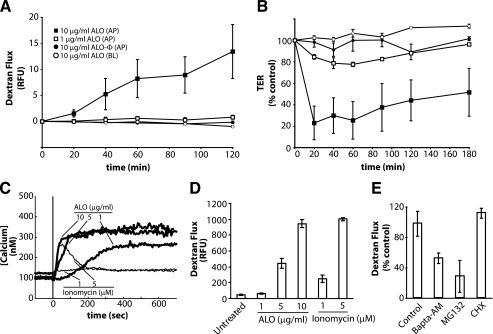
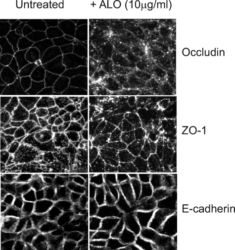
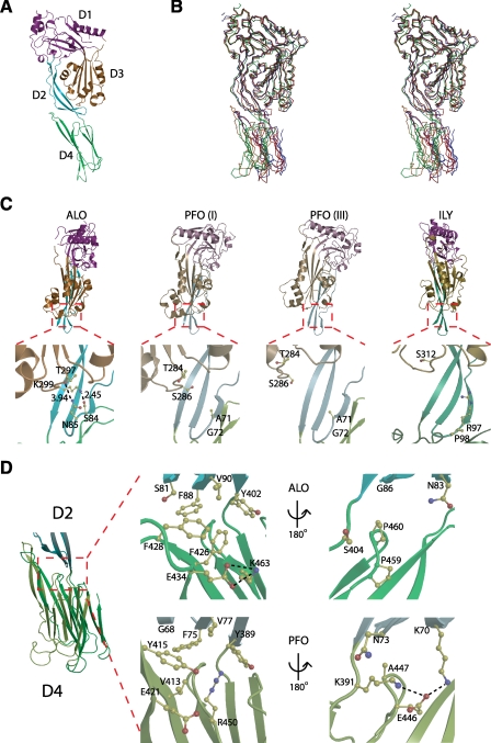
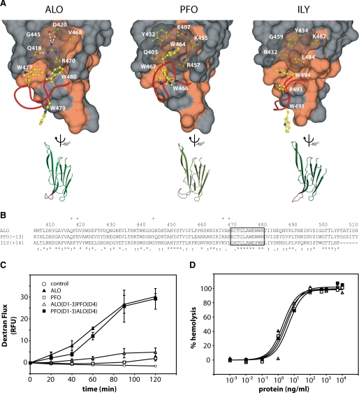
Anthrolysin O (ALO) is a pore-forming, cholesterol-dependent cytolysin (CDC) secreted by Bacillus anthracis, the etiologic agent for anthrax. Growing evidence suggests the involvement of ALO in anthrax pathogenesis. Here, we show that the apical application of ALO decreases the barrier function of human polarized epithelial cells as well as increases intracellular calcium and the internalization of the tight junction protein occludin. Using pharmacological agents, we also found that barrier function disruption requires increased intracellular calcium and protein degradation. We also report a crystal structure of the soluble state of ALO. Based on our analytical ultracentrifugation and light scattering studies, ALO exists as a monomer. Our ALO structure provides the molecular basis as to how ALO is locked in a monomeric state, in contrast to other CDCs that undergo antiparallel dimerization or higher order oligomerization in solution. ALO has four domains and is globally similar to perfringolysin O (PFO) and intermedilysin (ILY), yet the highly conserved undecapeptide region in domain 4 (D4) adopts a completely different conformation in all three CDCs. Consistent with the differences within D4 and at the D2-D4 interface, we found that ALO D4 plays a key role in affecting the barrier function of C2BBE cells, whereas PFO domain 4 cannot substitute for this role. Novel structural elements and unique cellular functions of ALO revealed by our studies provide new insight into the molecular basis for the diverse nature of the CDC family.


Figures

FIGURE 1.
FIGURE 1.
ALO induces C2BBE monolayer permeability. A, 3-kDa dextran-fluorescein flux in C2BBE monolayers treated apically (AP) or basolaterally (BL) with ALO or heat-inactivated ALO (ALO-Φ) at 1 μg/ml or 10 μg/ml. Flux is quantified by basolateral relative fluorescence units (RFU). B, transepithelial resistance of C2BBE monolayers treated with ALO at 1 or 10 μg/ml. Symbols represent the same conditions as seen in A. C, intracellular Ca2+ levels in C2BBE monolayers during ALO (thick lines) or ionomycin (thin lines) treatment. The monolayers were perfused with HBSS (which contains 1.26 mm Ca2+) and treated with either ALO or ionomycin at the indicated concentrations at time = 0 s (vertical line). D, dextran-fluorescein flux across C2BBE monolayers treated apically with ALO (0, 5, or 10 μg/ml) or ionomycin (1 or 5 μm) for 2 h. E, ALO (1 μg/ml) induced dextran-fluorescein flux across the C2BBE monolayer after pretreatment of C2BBE monolayers with either the proteasome inhibitor, MG132 (10 μm), for 2 h or BAPTA-AM (10 μm) for a 30-min pretreatment and 2-h treatment. Error bars, S.E. CHX, cycloheximide.
FIGURE 2.
FIGURE 2.
ALO induces alteration to occludin localization. As visualized by confocal immunofluorescence microscopy, the location of three junctional proteins was observed both prior to (left column) and after ALO (10 μg/ml) treatment for 2 h (right column). Occludin labeling (top panel) of untreated C2BBE monolayers revealed continuous staining around the cell periphery. When C2BBE monolayers are treated with ALO (10 μg/ml), occludin becomes internalized as granular intracellular puncta, and gaps within the continuous cell periphery localization become apparent. Another tight junction protein, ZO-1 (middle), shows similar peripheral and intracellular localization both prior to and after ALO treatment. E-cadherin (bottom), an adherens junction protein not associated with occludin or ZO-1, remains on the cell periphery both prior to and after ALO treatment.
FIGURE 3.
FIGURE 3.
Structural comparison of ALO with PFO (crystal form I), PFO (crystal form III), and ILY. A, side view of ALO colored by domain. D1 is colored magenta, D2 is colored cyan, D3 is colored orange, and D4 is colored green. B, stereo view of ALO aligned with PFO (I), PFO (III), and ILY using LSQ superimposition in COOT on all Cα′ atoms in D1 and D3 (70). ALO is colored red, PFO (I) is colored orange, PFO (III) is colored blue, and ILY is colored green. C, front view of ALO, PFO (I), PFO (III), and ILY aligned by D2. D4 has been omitted for clarity. Lower panels highlight the interactions or lack there of between the β-turn of D2 and transmembrane hairpin 2 of D3. D, side view of ALO and PFO (I) aligned by D2. The center panels show a close-up of the hydrophobic networks at the D2-D4 interface. The right panels show the interactions on the flip side of the D2-D4 interface.
FIGURE 4.
FIGURE 4.
Oligomerization analysis and hypothetical dimer configurations. A, chromatograms of ALO from S200 size exclusion chromatography coupled to differential pressure transducers and SLS and UV detectors. Data lines show UV (solid), RALS (dot-dash), LALS (dot-dot-dash), and DP (dotted). B, molecular weight (MW) and intrinsic viscosity (IV) patterns of ALO. Data lines show molecular weight (solid), LALS (dot-dot-dash), and IV (dotted). The retention volume of ALO (22.2 ml) is probably due to interactions with the column media. Using standard proteins for calibration, such a retention volume corresponds to a 15-20-kDa protein (not shown). Hence, measurement of molecular mass by SEC-SLS, which is independent from the retention volume on SEC, is appropriate for ALO. C, sedimentation velocity analysis of ALO from analytical ultracentrifugation at three different pH values: pH 5.5 (dashed), pH 7.0 (solid), and pH 8.0 (dotted). D, potential dimer interactions. Crystallographic dimers are found in ALO and PFO (I) crystals, and noncrystallographic dimers are seen in PFO (III) and ILY crystals. Domains 1-3 are colored blue, and D4 is colored orange.
FIGURE 5.
FIGURE 5.
Structural comparison of domain 4 and functional results from domain swap experiments. A, on the top are surface representations of D4. The red ribbon represents the main chain of the highly conserved undecapeptide region. The Trp side chains of the undecapeptide region are shown in yellow. The side chains under the translucent surface are thought to play a role in the orientation of the Trp side chains. Hydrophobic surfaces are colored coral. On the bottom are representations of D4 aligned with each other. The undecapeptide region is again highlighted in red. B, sequence alignment of D4s using ClustalW. Plus signs, residues not in the undecapeptide that are displayed in A. Asterisks, identical residues. Colons, conserved substitutions. Periods, semiconserved substitutions. C, 3-kDa dextran-fluorescein flux in C2BBE monolayers treated apically at 10 μg/ml. D, hemolysis of human erythrocytes after a 30-min treatment. RFU, relative fluorescence units.

References

    1. Rosado, C. J., Buckle, A. M., Law, R. H., Butcher, R. E., Kan, W. T., Bird, C. H., Ung, K., Browne, K. A., Baran, K., Bashtannyk-Puhalovich, T. A., Faux, N. G., Wong, W., Porter, C. J., Pike, R. N., Ellisdon, A. M., Pearce, M. C., Bottomley, S. P., Emsley, J., Smith, A. I., Rossjohn, J., Hartland, E. L., Voskoboinik, I., Trapani, J. A., Bird, P. I., Dunstone, M. A., and Whisstock, J. C. (2007) Science 317 1548-1551 - PubMed
    1. Rosado, C. J., Kondos, S., Bull, T. E., Kuiper, M. J., Law, R. H., Buckle, A. M., Voskoboinik, I., Bird, P. I., Trapani, J. A., Whisstock, J. C., and Dunstone, M. A. (2008) Cell. Microbiol. 10 1765-1774 - PMC - PubMed
    1. Giddings, K. S., Johnson, A. E., and Tweten, R. K. (2003) Proc. Natl. Acad. Sci. U. S. A. 100 11315-11320 - PMC - PubMed
    1. Giddings, K. S., Zhao, J., Sims, P. J., and Tweten, R. K. (2004) Nat. Struct. Mol. Biol. 11 1173-1178 - PubMed
    1. Tweten, R. K. (2005) Infect. Immun. 73 6199-6209 - PMC - PubMed

Publication types

MeSH terms